
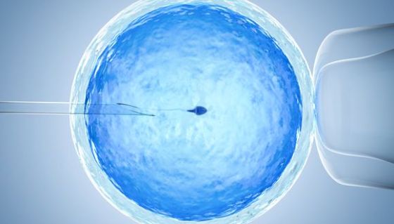
Accurate 3D imaging could significantly improve IVF treatments
Written on | Medicine
New Tel Aviv University technology allows clinicians to identify and select better-quality sperm, potentially increasing chances of pregnancy
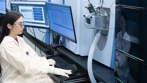
Tel Aviv University researchers have developed a safe and accurate 3D imaging method to identify sperm cells moving at a high speed.
The research was led by Prof. Natan Shaked of the Department of Biomedical Engineering at TAU’s Faculty of Engineering together with TAU doctoral student Gili Dardikman-Yoffe. The new technology could provide doctors with the ability to select the highest-quality sperm for injection into an egg during IVF treatment, potentially increasing a woman’s chance of becoming pregnant and giving birth to a healthy baby.
“The IVF procedure was invented to help fertility problems,” explains Prof. Shaked. “The most common type of IVF today is intra-cytoplasmic sperm injection (ICSI), which involves sperm selection by a clinical embryologist and injection into the woman’s egg. To that end, an effort is made to select the sperm cell that is most likely to create a healthy embryo.”
Choosing the right cells to make a baby
Under natural fertilization in the woman’s body, the fastest sperm to reach an egg is supposed to bear high-quality genetic material. Progressive movement allows this “best” sperm to overcome the veritable obstacle course of a woman’s reproductive system. “But this ‘natural selection’ is not available to the embryologist, who selects a sperm and injects it into the egg,” Prof. Shaked says. “Sperm cells not only move fast, they are also mostly transparent under regular light microscopy, and cell staining is not allowed in human IVF. Existing imaging technology that can examine the quality of the sperm’s genetic material may cause embryonic damage, so that too is prohibited. In the absence of more precise criteria, sperm cells are selected primarily according to external characteristics and their motility while swimming in water in a dish, which is very different from the natural environment of a woman’s body. “In our study, we sought to develop an entirely new type of imaging technology that would provide as much information as possible about individual sperm cells, does not require cell staining to enhance contrast, and has the potential for enabling the selection of optimal sperm in fertilization treatments.”A hologram of sperm cells
The researchers chose light computed tomography (CT) technology for the unique task of sperm cell imaging. “In a standard medical CT scan, the device rotates around the subject and sends out X-rays that produce multiple projections, ultimately creating a 3D image of the body,” says Prof. Shaked. “In the case of the sperm, instead of rotating the device around this tiny subject, we relied on a natural feature of the sperm itself: Its head is constantly rotating during the forward movement. We used weak light (and not X-rays), which does not damage the cell. We recorded a hologram of the sperm cell during ultrafast movement and identified various internal components according to their refractive index. This creates an accurate, highly dynamic 3D map of its contents without using cell staining.” Using this technique, the researchers obtained a clear and accurate CT image of the sperm at very high resolution in four dimensions: three dimensions in the space at resolution of less than half a micron (one micron equals one millionth of a meter) and the exact time (motion) dimension of the second sub-millisecond. “Our new development provides a comprehensive solution to many known problems of sperm imaging,” Prof. Shaked says. “We were able to create high-resolution imaging of the sperm head while it was moving fast, without the need for stains that could harm the embryo. The new technology can greatly improve the selection of sperm cells in vitro, potentially increasing the chance of pregnancy and the birth of a healthy baby. “To help diagnose male fertility problems, we intend to use our new technique to shed light on the relationship between the 3D movement, structure and contents of sperm and its ability to fertilize an egg and produce a viable pregnancy,” Prof. Shaked concludes. “We believe that such imaging capabilities will contribute to other medical applications, such as developing efficient biomimetic micro-robots to carry drugs within the body.”Related posts
Heart Disease’s Cancer Link Unveiled

Do Green Environments Help Heart Patients Live Longer?

TAU Receives $12.67M Grant for Medical Simulation Center

Breaking the MedTech Glass Ceiling

Summer Glow: How Sun Exposure Boosts Fertility in Women Ages 30-40

Are We Close to Ending Alzheimer’s Memory Loss?

Destroying Cancer: new drug delivery system containing RNA therapy can target cancer cells in bone marrow
Stress Makes Vaccines Less Effective
Researchers Induce Cancer Cell “Suicide”
Metabolomics – A New Frontier in Preventive Medicine

Older Bats do Suffer from Age-related Hearing Loss

Operation Guardian of the Walls: Women, Young People and Residents of the South Paid the Heaviest Price
Discovery May Lead to Personalized Medicine for Infectious Diseases

One Third of Normal-Weight Individuals are Obese

Breakthrough Gene Therapy Offers Hope for Severe Developmental Epilepsy in Children
