Author: AUFTAU

The Silent Prophets

TAU researchers prove that silent mutations can predict development of cancer cells.

Our genome, our complete set of genetic instructions, contains mutations that can change the sequence of amino acids in the coded proteins. Since these proteins are responsible for the various cell mechanisms, such mutations are involved in turning healthy cells into cancer cells. In contrast, there are so-called ‘silent mutations’ that don’t change the sequence of amino acids in proteins. In recent years, it has been shown that silent mutations, both in and out of the cell’s genetic coding region, can affect gene expression, and may be associated with the development and spread of cancer cells. However, the question of whether silent mutations can help identify cancer types or predict patients’ chances of survival has never before been investigated with quantitative tools. Researchers from TAU’s Department of Biomedical Engineering and the Zimin Institute for Engineering Solutions Advancing Better Lives have been able to predict both the type of cancer and patients’ survival probability based on silent mutations in cancer genomes – a proof of concept that may well save lives in the future.

Predictive Power Similar to That of ‘Ordinary’ Mutations.

The groundbreaking study, led by Prof. Tamir Tuller and research student Tal Gutman, is based on about three million mutations from cancer genomes of 9,915 patients. The researchers attempted to identify the type of cancer and predict survival probability 10 years after the initial diagnosis – on the basis of silent mutations alone. They found that the predictive power of silent mutations is often similar to that of ‘ordinary’, non-silent mutations.

In addition, they discovered that by combining information from silent and non-silent mutations classification could be improved for 68% of the cancer types, and the best survival estimations could be obtained up to nine years after diagnosis. In some types of cancer classification was improved by up to 17%, while prognosis was improved by up to 5%. The findings of the study were recently published in NPJ Genomic Medicine.

Silent, Yet Making Noise

“‘Silent mutations’ have been ignored by researchers for many years,” explains Prof. Tuller. “In our study, about 10,000 cancer genomes of every type were analyzed, demonstrating for the first time that silent mutations do have diagnostic value – for identifying the type of cancer, as well as prognostic value – for predicting how long the patient is likely to survive.”

According to the professor, the cell’s genetic material holds two types of information: first, the sequence of amino acids to be produced, and second, when and how much to produce of each protein – namely regulation of the production process. “Even if they don’t change the structure of the protein, silent mutations can influence the process of protein production (gene expression), which is just as important. If a cell prodces much smaller quantities of a certain protein – it’s almost as though the protein has been eliminated altogether.”

“Another important aspect, which can also be affected by silent mutations, is the protein’s 3D folding, which impacts its functions: Proteins are long molecules usually consisting of many hundreds of amino acids, and their folding process begins when they are produced in the ribosome. Folding can be affected by the rate at which the protein is produced, which may in turn be affected by silent mutations.”

“Also, in some cases, silent mutations can impact a process called splicing, in which pieces of the genetic material are cut and rearranged to create the final sequence in the protein.”

Apparently, silent mutations can actually make a lot of noise, and Prof. Tuller and his colleagues were able to quantify their impact for the first time.

Saving as Many Lives as Possible

To test their hypothesis and quantify the effect of the silent mutations, the researchers used public genetic information about cancer genomes from the NIH in the USA. Applying machine learning techniques to this data, the team obtained predictions of the type of cancer and prognoses for patients’ survival – based on silent mutations alone. They then compared their results with real data from the database.

“The results of our study have several important implications,” says Prof. Tuller. “First of all, there is no doubt that by using silent mutations we can improve existing diagnostic and prognostic models. It should be noted that even a 17% improvement is very significant, because there are real people behind these numbers – sometimes even ourselves or our loved ones.”

“Doctors discovering metastases would like to know where they came from and how the disease has developed, in order to prescribe the best treatment. If, hypothetically, instead of giving wrong diagnoses and prognostics to five out of ten cancer patients, they only make mistakes in four out of ten cases, millions of lives may ultimately be saved. In addition, our results indicate that in many cases silent mutations can by themselves provide predictive power that is similar to that of non-silent mutations. These results are especially significant for a range of technologies currently under development, striving to diagnose cancer types based on DNA from malignant sources identified in simple blood tests. Since most of our DNA does not code for proteins, we may assume that most cancer DNA obtained from blood samples will contain silent mutations.”

The new study has implications for all areas of oncological research and treatment. Following this proof of concept, the researchers intend to establish a startup with Sanara Ventures, focusing on silent mutations as a diagnostic and prognostic tool.

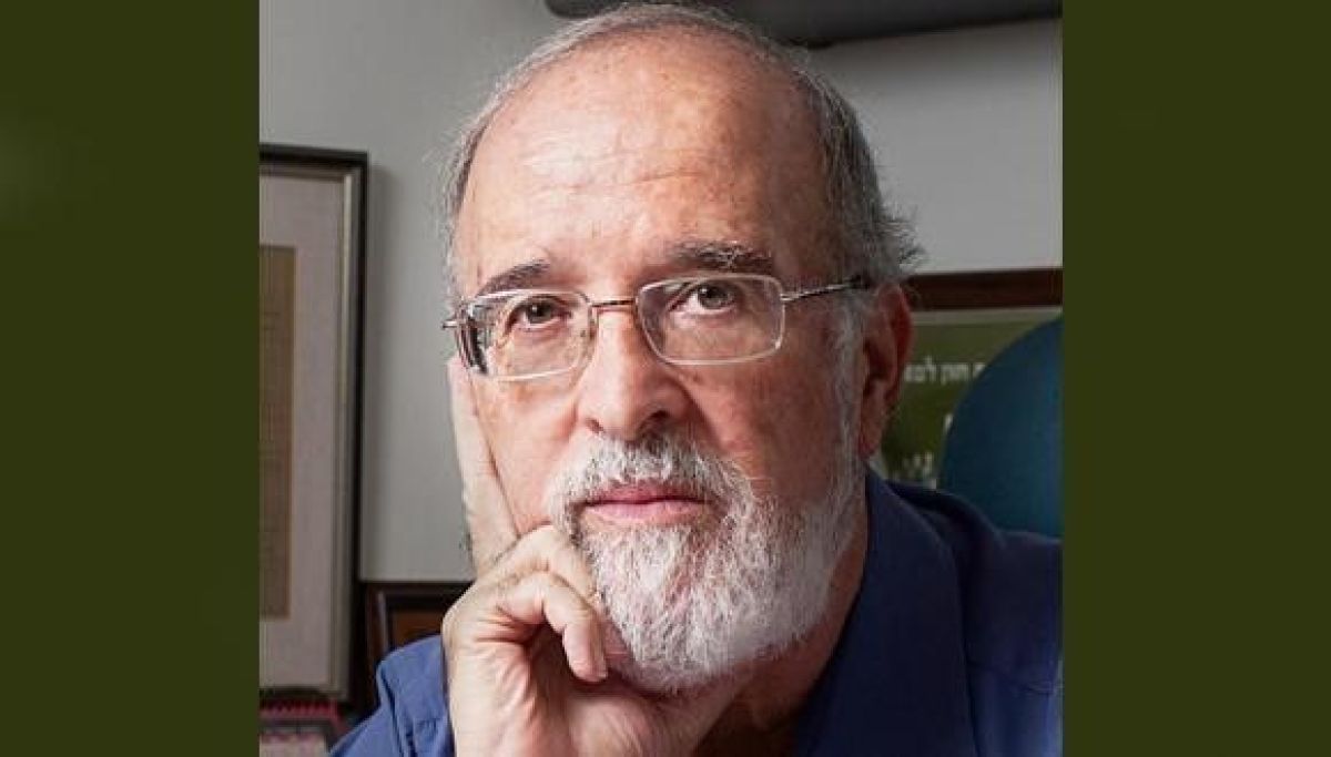
Featured image: Prof. Tamir Tuller (Photo: Rafael Ben Menashe)

Want to Fall in Love? Step Outside in The Sun

Exposure to ultraviolet radiation from sunlight enhances romantic passion in humans.

Any Tel Avivian will tell you that the perfect place for a first date is at the beach. Now, we have the science to support that claim. Researchers at Tel Aviv University have found that exposure to ultraviolet radiation from sunlight enhances romantic passion in humans. In the study, men and women were exposed to UVB (ultraviolet radiation type B) under controlled conditions, and the findings were unequivocal: increased levels of romantic passion in both genders.

Sun + Skin = Love

The study revealed that exposure to sunlight affects the regulation of the endocrine system responsible for the release of sexual hormones in humans. The discovery may lead to practical applications down the line, such as UVB treatments for sexual hormone disorders.

In animal models, the effect was dramatic: the females’ hormone levels rose significantly, enlarging their ovaries and prolonging their mating season; the attraction between males and females increased; and both were more willing to engage in sexual intercourse.

The researchers repeated the experiment on the animal model, this time removing from the skin a protein called p53, which identifies DNA damage and activates pigmentation during exposure to sunlight as protection against its adverse effects. The removal of the protein eliminated the effect of UVB exposure on the animals’ sexual behavior, convincing the researchers that exposure to radiation through the skin was the cause of the observed hormonal, physiological and behavioral changes, and that the protective system is also responsible for the regulation of sexuality.

Furless Humans and Sun Exposure

In the 32 human subjects of the study, all treated with UVB phototherapy at the Tel Aviv Sourasky (Ichilov) and Assuta Medical Centers, both genders exhibited a rise in romantic passion, and males also noted an increase in levels of aggression.

Similar results were found when the subjects were asked to avoid sunlight for two days, and then tan themselves for approximately 25 minutes. Blood tests revealed that exposure to sunlight resulted in a higher release of hormones like testosterone compared to one day before exposure. A rise in testosterone in males during the summer was also found in analyses of data from the Israeli health maintenance organizations Clalit and Maccabi Health Services.


Prof. Carmit Levy (on the left) & PhD student Roma Parikh.

The new discovery from TAU may lead to future practical applications, such as UVB treatments for sexual hormone disorders. The breakthrough opens up for further discoveries in basic science, “As humans, we have no fur, and our skin is thus directly exposed to sunlight. We are only beginning to understand what this exposure does to us, and the key roles it might play in various physiological and behavioral processes. It’s only the tip of the iceberg,” says Prof. Carmit Levy from the Department of Human Molecular Genetics and Biochemistry at the Sackler Faculty of Medicine.

The study was led by PhD student Roma Parikh and Ashchar Sorek from the laboratory of Prof. Levy. UVB phototherapy was administered to the subjects at the Tel Aviv Sourasky (Ichilov) and Assuta Medical Centers. The groundbreaking discovery was published as a cover story in the prestigious scientific journal Cell Reports

Recordings of the magnetic field from 9,000 years ago teach us about the magnetic field today

View west of the 1999 excavations, Stratum IIB,  Tel Tifdan/ Wadi Fidan. Photo courtesy of Thomas E. Levy

Tel Aviv University Research Links Archaeology, Physics, and Geophysics

  • Burnt archaeological flints enable us to determine the strength of the Earth’s magnetic field during prehistoric periods.
  • Information about the magnetic field in antiquity helps us understand the magnetic field today. Researchers: “the current weakening of the field is a reversible trend; Seven thousand six hundred years ago, the strength of the magnetic field was even lower than today, but within approximately 600 years, it gained strength and again rose to high levels.”

International research by Tel Aviv University, the Istituto Nazionale di Geofisica e Vulcanologia, Rome, and the University of California San Diego uncovered findings regarding the magnetic field that prevailed in the Middle East between approximately 10,000 and 8,000 years ago. Researchers examined pottery and burnt flints from archaeological sites in Jordan, on which the magnetic field during that time period was recorded. Information about the magnetic field during prehistoric times can affect our understanding of the magnetic field today, which has been showing a weakening trend that has been cause for concern among climate and environmental researchers.

The research was conducted under the leadership of Prof. Erez Ben-Yosef of the Jacob M. Alkow Department of Archaeology and Ancient Near Eastern Cultures at Tel Aviv University and Prof. Lisa Tauxe, head of the Paleomagnetic Laboratory at the Scripps Institution of Oceanography, in collaboration with other researchers from the University of California at San Diego, Rome and Jordan. The article was published in the journal PNAS.

Prof. Erez Ben-Yosef
Photo courtesy of Yoram Reshef

“Albert Einstein characterized the planet’s magnetic field as one of the five greatest mysteries of modern physics…”

Prof. Ben-Yosef explains, “Albert Einstein characterized the planet’s magnetic field as one of the five greatest mysteries of modern physics. As of now, we know a number of basic facts about it: The magnetic field is generated by processes that take place below a depth of approximately 3,000 km beneath the surface of the planet (for the sake of comparison, the deepest human drilling has reached a depth of only 20 km); it protects the planet from the continued bombardment by cosmic radiation and thus allows life as we know it to exist; it is volatile and its strength and direction are constantly shifting, and it is connected to various phenomena in the atmosphere and the planet’s ecological system, including – possibly – having a certain impact on climate. Nevertheless, the magnetic field’s essence and origins have remained largely unresolved. In our research, we sought to open a peephole into this great riddle.”

The researchers explain that instruments for measuring the strength of the Earth’s magnetic field were first invented only approximately 200 years ago. In order to examine the history of the field during earlier periods, science is helped by archaeological and geological materials that recorded the properties of the field when they were heated to high temperatures. The magnetic information remains “frozen” (forever or until another heating event) within tiny crystals of ferromagnetic minerals, from which it can be extracted using a series of experiments in the magnetics laboratory. Basalt from volcanic eruptions or ceramics fired in a kiln are frequent materials used for these types of experiments. The great advantage in using archaeological materials as opposed to geological is the time resolution: While in geology dating is on the scale of thousands years at best, in archaeology the artifacts and the magnetic field that they have recorded can be dated at a resolution of hundreds and sometimes even tens of years (and in specific cases, such as a known destruction event, even give an exact date). The obvious disadvantage of archaeology is the young age of the relevant artifacts: Ceramics, which have been used for this purpose up until now, were only invented 8,500 years ago.
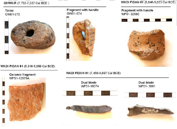
Burnt flints and ceramics used to reconstruct the strength of the ancient geomagnetic field
(https://doi.org/10.1073/pnas.2100995118)

The current study is based on materials from four archaeological sites in Wadi Feinan (Jordan), which have been dated (using carbon-14) to the Neolithic period – approximately 10,000 to 8,000 years ago – some of which predate the invention of ceramics. Researchers examined the magnetic field that was recorded in 129 items found in these excavations, and this time, burnt flint tools were added to the ceramic shards.  Prof. Ben-Yosef: “This is the first time that burnt flints from prehistoric sites are being used to reconstruct the magnetic field from their time period. About a year ago, groundbreaking research at the Hebrew University was published, showing the feasibility of working with such materials, and we took that one step forward, extracting geomagnetic information from tightly dated burned flint. Working with this material extends the research possibilities tens of thousands of years back, as humans used flint tools for a very long period of time prior to the invention of ceramics. Additionally, after enough information is collected about the changes in the geomagnetic field over the course of time, we will be able to use it in order to date archaeological remains”.

Wadi Fidan 61 Pottery Neolithic.
Photo courtesy of Thomas E. Levy

An additional and important finding of this study is the strength of the magnetic field during the time period that was examined. The archaeological artifacts demonstrated that at a certain stage during the Neolithic period, the field became very weak (among the weakest values ever recorded for the last 10,000 years), but recovered and strengthened within a relatively short amount of time. According to Prof. Tauxe, this finding is significant for us today: “In our time, since measurements began less than 200 years ago, we have seen a continuous decrease in the field’s strength. This fact gives rise to a concern that we could completely lose the magnetic field that protects us against cosmic radiation and therefore, is essential to the existence of life on Earth. The findings of our study can be reassuring: This has already happened in the past. Approximately 7,600 years ago, the strength of the magnetic field was even lower than today, but within approximately 600 years, it gained strength and again rose to high levels.”

The research was carried out with the support of the US-Israel Binational Science Foundation, which encourages academic collaborations between universities in Israel and in the US. The researchers note that in this case, the collaboration was particularly essential to the success of the study because it is based on a tight integration of methods from the fields of archaeology and geophysics, and the insights that were obtained are notably relevant to both of these disciplines.

Link to the article:

https://doi.org/10.1073/pnas.2100995118

Continue reading

First 3D-bioprinting of entire active tumor

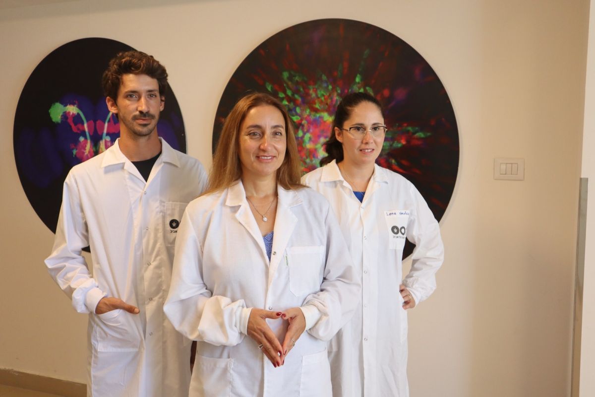
Research team(Left to right): Eilam Yeini, Prof. Satchi-Fainaro and Lena Neufeld

Scientific breakthrough in the battle against cancer

The 3D print of glioblastoma – the deadliest type of brain cancer – is printed from human glioblastoma tissues containing all components of the malignant tumor

Researchers: the breakthrough will enable much faster prediction of best treatments for patients, accelerate the development of new drugs and discovery of new druggable targets

A scientific achievement for researchers at Tel Aviv University: printing an entire active and viable glioblastoma tumor using a 3D printer. The 3D-bioprinted tumor includes a complex system of blood vessel-like tubes through which blood cells and drugs can flow, simulating a real tumor.

Illustration.
Credit: Veronica Hughes, PhD of STEAM visuals

The study was led by Prof. Ronit Satchi-Fainaro, Sackler Faculty of Medicine and Sagol School of Neuroscience, Director of the Cancer Biology Research Center, Head of the Cancer Research and Nanomedicine Laboratory and Director of the Morris Kahn 3D-BioPrinting for Cancer Research Initiative, at Tel Aviv University.

The new technology was developed by PhD student Lena Neufeld, together with other researchers at Prof. Satchi-Fainaro’s laboratory:  Eilam Yeini, Noa Reisman, Yael Shtilerman, Dr. Dikla Ben-Shushan, Sabina Pozzi, Dr. Galia Tiram, Dr. Anat Eldar-Boock and Dr. Shiran Farber.  

The 3D-bioprinted models are based on samples from patients, taken directly from operating rooms at the Tel Aviv Sourasky Medical Center. The new study’s results were published today in the prestigious journal Science Advances.

“Glioblastoma is the most lethal cancer of the central nervous system, accounting for most brain malignancies”

“Glioblastoma is the most lethal cancer of the central nervous system, accounting for most brain malignancies,” says Prof. Satchi-Fainaro. “In a previous study, we identified a protein called P-Selectin, produced when glioblastoma cancer cells encounter microglia – cells of the brain’s immune system. We found that this protein is responsible for a failure in the microglia, causing them to support rather than attack the deadly cancer cells, helping the cancer spread. However, we identified the protein in tumors removed during surgery, but not in glioblastoma cells grown on 2D plastic petri dishes in our lab. The reason is that cancer, like all tissues, behaves very differently on a plastic surface than it does in the human body. Approximately 90% of all experimental drugs fail at the clinical stage because the success achieved in the lab is not reproduced in patients.”

Prof. Ronit Satchi-Fainaro

To address this problem, the research team led by Prof. Satchi-Fainaro and PhD student Lena Neufeld, recipient of the prestigious Dan David Fellowship, created the first 3D-bioprinted model of a glioblastoma tumor, which includes 3D cancer tissue surrounded by extracellular matrix, which communicates with its microenvironment via functional blood vessels.

Microscopic image of the 3D-bioprinted glioblastoma model. The bioprinted blood vessels are covered with endothelial cells (red) and pericytes (cyan). The blood vessels are surrounded with a brain-mimicking tissue composed of gliblastoma cells (blue) and the brain microenvironment cells (green). Different drugs or cells can be perfused through the 3D-bioprinted blood vessels to test their effect on the tumor tissue

“It’s not only the cancer cells…”

“It’s not only the cancer cells,” explains Prof. Satchi-Fainaro. “It’s also the cells of the microenvironment in the brain; the astrocytes, microglia and blood vessels connected to a microfluidic system – namely a system enabling us to deliver substances like blood cells and drugs to the tumor replica. Each model is printed in a bioreactor we have designed in the lab, using a hydrogel sampled and reproduced from the extracellular matrix taken from the patient, thereby simulating the tissue itself. The physical and mechanical properties of the brain are different from those of other organs, like the skin, breast, or bone. Breast tissue consists mostly of fat, bone tissue is mostly calcium; each tissue has its own properties, which affect the behavior of cancer cells and how they respond to medications. Growing all types of cancer on identical plastic surfaces is not an optimal simulation of the clinical setting.”

After successfully printing the 3D tumor, Prof. Satchi-Fainaro and her colleagues demonstrated that unlike cancer cells growing on petri dishes, the 3D-bioprinted model has the potential to be effective for rapid, robust, and reproducible prediction of the most suitable treatment for a specific patient.

“We proved that our 3D model is better suited for prediction of treatment efficacy, target discovery and drug development in three different ways.

First, we tested a substance that inhibited the protein we had recently discovered, P-Selectin, in glioblastoma cell cultures grown on 2D petri dishes, and found no difference in cell division and migration between the treated cells and the control cells which received no treatment. In contrast, in both animal models and in the 3D-bioprinted models, we were able to delay the growth and invasion of glioblastoma by blocking the P-Selectin protein.

This experiment showed us why potentially effective drugs rarely reach the clinic simply because they fail tests in 2D models, and vice versa: why drugs considered a phenomenal success in the lab, ultimately fail in clinical trials. In addition, collaborating with the lab of Dr. Asaf Madi of the Department of Pathology at TAU’s Faculty of Medicine, we conducted genetic sequencing of the cancer cells grown in the 3D-bioprinted model, and compared them to both cancer cells grown on 2D plastic and cancer cells taken from patients.

Thus, we demonstrated a much greater resemblance between the 3D-bioprinted tumors and patient-derived glioblastoma cells grown together with brain stromal cells in their natural environment. Through time, the cancer cells grown on plastic changed considerably, finally losing any resemblance to the cancer cells in the patient’s brain tumor sample.

The third proof was obtained by measuring the tumor growth rate. Glioblastoma is an aggressive disease partially because it is unpredictable: when the heterogeneous cancer cells are injected separately into model animals, the cancer will remain dormant in some, while in others, an active tumor will develop rapidly. This makes sense because we, as humans, can die peacefully of old age without ever knowing we have harbored such dormant tumors. On the dish in the lab, however, all tumors grow at the same rate and spread in the same rate. In our 3D-bioprinted tumor, the heterogeneity is maintained and development is similar to the broad spectrum that we see in patients or animal models.”

“…perhaps the most exciting aspect is finding novel druggable target proteins and genes in cancer cells…”

According to Prof. Satchi-Fainaro, this innovative approach will also enable the development of new drugs, as well as discovery of new drug targets – at a much faster rate than today. Hopefully, in the future, this technology will facilitate personalized medicine for patients.

“If we take a sample from a patient’s tissue, together with its extracellular matrix, we can 3D-bioprint from this sample 100 tiny tumors and test many different drugs in various combinations to discover the optimal treatment for this specific tumor. Alternately, we can test numerous compounds on a 3D-bioprinted tumor and decide which is most promising for further development and investment as a potential drug.

But perhaps the most exciting aspect is finding novel druggable target proteins and genes in cancer cells – a very difficult task when the tumor is inside the brain of a human patient or model animal. Our innovation gives us unprecedented access, with no time limits, to 3D tumors mimicking better the clinical scenario, enabling optimal investigation.”

Illustration for demonstration of 3D printing of a tumor in a brain Microenvironment according to a computed 3D model

The study was funded by the Morris Kahn Foundation, European Research Council (ERC), Israel Cancer Research Fund (ICRF), the Israel Cancer Association and Israel Science Foundation (ISF), and Check Point Software Technologies LTD.

Continue reading

Prof. Isaac Ben-Israel Receives the IEEE Leadership Award

For his outstanding leadership contributions to homeland security, cyber security and resilience.

The Institute of Electrical and Electronics Engineers (IEEE) has extended its IEEE TCHS Outstanding Leadership Award in the field of cyber for the year 2021 to Major Gen. (Ret.) Prof. Isaac Ben-Israel, Head of the ICRC – Blavatnik Interdisciplinary Cyber Research Center.

The announcement of IEEE’s award committee members: “This award recognizes an individual’s outstanding leadership contributions to the homeland security community in general and to the field of cyber security and resilience in particular. The award recognizes the leadership and visionary contributions to the society at large through the promotion and applications of security and resilience concepts to a variety of technology, science, and business domains.”

“It is a great honor and I am very excited to receive the IEEE Award. I see great importance in receiving the award not only personally but also nationally, as the award is not only a recognition of my work but a recognition of Israel’s central and unique status in the cyber world,” said Prof. Isaac Ben-Israel.

As part of his wide-ranging activities, Prof. Ben Israel headed the project to formulate Israel’s national cyber program, and he heads the anual “Cyber ​​Week” events, one of the world’s leading cybersecurity conferences, where experts from industry, government and academia across the globe come together to exchange cyber dialogue on current issues, trends and technological solutions.

In 2012, Prof. Ben Israel was elected a Lifetime Member of the International Academy of Austronautics (IAC) and became a member of the Research, Innovation and Enterprise Council (RIEC) in Singapore. In addition to his academic work, Prof. Ben-Israel serves as Chairman of Israel Space Agency and he has received the Israel Defense Forces Award twice.

IEEE is the world’s largest technical professional organization (419,000 members from over 160 countries) dedicated to advancing technology for the benefit of humanity. 

Where Have All the Birds Gone?

Humans Behind Extinction of Hundreds of Bird Species Over the Last 50,000 Years.

A new study from Tel Aviv University and the Weizmann Institute revealed that over the last 20,000-50,000 years, birds have undergone a major extinction event, inflicted chiefly by humans, which caused the disappearance of about 10%-20% of all avian species. The vast majority of the extinct species shared several features: they were large, they lived on islands, and many of them were flightless.

The main cause for extinction of species by humans today has evolved from being hunting to the destruction of the animals’ natural habitats, but the researchers hope their findings will serve as warning signals regarding bird species currently threatened with extinction.

The study was led by Prof. Shai Meiri of the School of Zoology at The George S. Wise Faculty of Life Sciences and the Steinhardt Museum of Natural History at Tel Aviv University, and Amir Fromm of the Weizmann Institute of Science. The paper was published in the Journal of Biogeography.

Human-Inflicted Extinction

Prof. Meiri: “We conducted a comprehensive review of scientific literature, and for the first time collected quantitative data on the numbers and traits of extinct species of birds worldwide. Those that became extinct in the last 300 years or so are relatively well known, while earlier species are known to science from remains found in archaeological and paleontological sites worldwide. Altogether we were able to list 469 avian species that became extinct over the last 50,000 years, but we believe that the real number is much higher.”

The researchers believe that the vast extinction was caused primarily by humans who hunted the birds for food, or by animals brought to islands by humans – that fed on the birds and/or their eggs. This assumption is based on the fact that the greater part of bird remains was found on human sites, apparently belonging to birds consumed by the inhabitants, and in most cases the extinctions occurred shortly after the arrival of humans.

Coveted Targets for Hunters

Most extinct species shared three major features:

  1. About 90% of them lived on islands – When humans arrived on the island, the birds were hunted by them, or fell victim to other animals introduced by humans, such as pigs, rats, monkeys, and cats.
  2. Most extinct bird species were large, some very large – The body mass of the extinct species was found to be up to 10 times as large as that of surviving species. The larger birds provided humans with a great quantity of food, thus they were a preferred target for hunters. Previous studies have found a similar phenomenon among mammals and reptiles, especially lizards and turtles that lived on islands: the larger ones were hunted by humans and became extinct.
  3. A large portion of the extinct bird species were flightless, and often unable to escape their pursuers – The study found that the number of flightless bird species that became extinct is double the number of flightless species still existing today; all in all, 68% of the flightless bird species known to science became extinct. One of the better-known examples is the moa bird in New Zealand: 11 species of moa became extinct within 300 hundred years, due to hunting by humans

Prof. Meiri: “Our study indicates that before the major extinction event of the past millennia, many more large, even giant, as well as flightless avian lived on our globe, and the diversity of birds living on islands was much greater than today. We hope that our findings can serve as warning signals regarding bird species currently threatened with extinction, and it is therefore important to check whether they have similar features. It must be noted, however, that conditions have changed considerably, and today the main cause for extinction of species by humans is not hunting but rather the destruction of natural habitats.”

Featured image: Bird species at the Zoological Garden

For the first time: The “God Particle” has been characterized in its decay into a pair of charm quarks

TAU researchers contribute further understanding of elusive elementary particle that gives mass to everything in the universe

Physicists worldwide have been captivated by the Higgs boson particle, also known as the “God Particle”. Its discovery a decade ago made waves in the physics community, and had researchers curious to learn more about its properties. TAU researchers have now succeeded, as part of a groundbreaking study, to describe a rare physical process through which the Higgs boson decays into a pair of rare elementary particles. The rate of this decay process can now be characterized more precisely and completely than before.

The new study was conducted as part of the ATLAS experiment at the Large Hadron Collider (LHC) at CERN (Geneva) by Prof. Erez Etzion and doctoral students Guy Koren, Hadar Cohen and David Reikher from the Raymond and Beverly Sackler School of Physics and Astronomy, Raymond and Beverly Sackler Faculty of Exact Sciences, at Tel Aviv University. It was a collaboration with the research team of Prof. Eilam Gross from the Weizmann Institute of Science and others.

Learning More About Forces in Nature

Over fifty years ago, physicists Prof. Peter Higgs and Prof. Francois Englert (who since 1984 has been a Sackler Fellow by special appointment in the TAU School of Physics and Astronomy) estimated that a new particle might exist whose field “provides the mass” to the elementary particles in our world.

In 2012, the end of a 30-year hunt for the Higgs boson was celebrated. Israeli researchers were senior partners in this discovery, and Prof. Halina Abramowicz, who was part of the TAU team, said “The discovery of the Higgs-like particle affirms the world view that the universe is made up of straightforward, symmetrical laws and that humans are the byproduct of disruptions in that symmetry.” Higgs and Englert won the Nobel Prize the following year.

The Challenge of Creating the Higgs boson 

In the particle accelerator, pairs of protons are made to collide with each other at extremely high velocities. In such energetic collisions, various interesting processes can occur, from which, one can learn about the nature of our universe. The way in which these processes are investigated, is by means of a complex array of particle detectors placed around the points of collision, enabling reconstruction of the types of particles that are generated during the collision, as well as their features. A vast range of processes can occur during the collisions, and each has its own unique “signature” in the detector. In order to extract rare events and acquire new insights about the elementary particles and forces in nature, large amounts of statistical data must be collected (i.e. a very large number of collisions must be observed).

The Higgs boson is, as mentioned, a relatively heavy elementary particle, but can be created in collision between protons, as long as the accelerator’s energy is high enough. Immediately after its creation, it decays into lighter particles.

“It is interesting to investigate into which types of particles the Higgs decays, and with what frequency it decays into each type of particle,” says Guy Koren. “To help answer that question, our group is trying to measure the rate at which the Higgs boson decays into particles called ‘charm quarks’.” Quarks are a specific type of particles that share similar features. They compound, for instance, the protons and neutrons, which are in the nuclei of atoms. Koren continues to explain that measuring the decay of Higgs boson into ‘charm quarks’ is not a simple mission, for two reasons: 1. Only one out of billions of collisions [between protons] result in the creation of Higgs bosons. Furthermore, only three percent of the Higgs bosons that do emerge proceed to decay into charm quarks. 2. Five additional types of quarks exist, and they all leave similar signatures in the detectors. So, even when the process does take place, it is very hard to identify.

More Information About The Rate of Decay 

Despite all the collisions that have been collected since 2012, the group from Tel Aviv has not yet identified enough decays of Higgs bosons into charm quarks to measure the rate of the process with the required statistical accuracy.

Nevertheless, sufficient data has been accumulated to state what the maximal rate of the process is with respect to the theoretical predictions. A rate of decay higher than the predicted rate would constitute a first important indicator for “new” physics or expansion of the currently accepted model – the standard model of elementary particles. From the current measurement, the researchers conclude (with a well-defined statistical certainty) that there is no chance that the rate of decay of the Higgs boson into charm quark is 8.5 (or more) times higher than the theoretical predictions, otherwise enough such decays would have been observed in order to measure it. “This is the first time that anyone has ever succeeded in saying something important about the rate of this specific decay based on a direct measurement of it, therefore it is a very important and significant statement in our field,” explains Koren

The research is not yet over, however. Higgs’ decays into quarks of smaller masses have yet to be observed. As a result, the researchers cannot be certain that the same ‘rules’ apply to quarks from those generations. “If it should appear that the Higgs boson decays at a rate that is not proportional to mass (squared) of the particles, there could be far-reaching implications for our understanding of the universe,” explains Prof. Etzion.

Featured image: Illustration: The European Organization for Nuclear Research (CERN)’s LHC accelerator, by which the Higgs boson was detected in 2012 in the ATLAS and CMS experiments

New Warning Sign for Breast Cancer

TAU-led research lays groundwork for preventive treatment that may save millions of lives.

A team led by Tel Aviv University identified a new indicator of metastatic breast cancer, laying the groundwork for preventive treatment that could save millions of lives.

Metastatic breast cancer, also known as Stage 4 breast cancer, occurs when cancer has spread, or “metastasized,” to other parts of the body. Mortality from breast cancer is almost exclusively a result of tumor metastasis, and lungs are one of the main metastatic sites. The five-year survival rate for women with metastatic breast cancer is estimated at 28%.

Investigating The “Black Box” of Breast Cancer 

“Breast cancer patients, as well as patients with many other types of cancer, do not die from the primary tumor, but from distant metastases which have developed, sometimes after years, in essential organs such as the lungs and brain,” said the study’s lead researcher, Prof. Neta Erez, Chair of the Department of Pathology at TAU’s Sackler Faculty of Medicine. “Understanding the body’s preparation for the reception of metastases at an early stage may save millions of lives.”

The researchers explain that metastases can appear several years after the initial cases are treated. Today, methods used for follow-up screening identify metastases only when they are quite large–when the disease is at an advanced stage and unlikely to be cured.

For this reason, Erez’s research group is investigating the black box—the time period between apparent recovery and the appearance of metastases to understand the metastatic process and to find ways of blocking it in early stages. Their research in recent years has revealed that certain tissues, in organs where the metastases are set to arrive, “prepare the area” for reception and produce a hospitable environment for them, a long time before the appearance of the metastases themselves. In the present study, the research team searched for signs of these changes, which may be used in the future to identify the start of the process that predicts metastases. The researchers identified these changes in the area known as “the micro-environment” of the tumor, and specifically in connective tissue known as fibroblasts which are found in the lungs among other places. 

“In a normal situation, fibroblasts play a central role in healing wounds and injury to the lungs, but recent studies revealed that cancer is successful in recruiting them and causing them to produce a supportive environment for it,” said Erez.

What is Happening in the Micro-environment of the Metastases?

The researchers compared genes sequenced from healthy lungs, from lungs with micro-metastases (very small metastases which cannot be identified using existing clinical tools) and from lungs with large metastases, in a state of advanced disease.

By identifying and comparing the respective development in the three different types of sample tissues, the researchers succeeded, for the first time, in characterizing the process that occurs in the micro-environment of the metastases. The findings provide valuable understanding about how cancer cells grow, which can then be leveraged for detection by existing imaging methods and treated to prevent metastasis.

The study’s leading research team from Erez’s laboratory included Dr. Ophir Shani and Dr. Yael Raz along with additional researchers from Tel Aviv University, Sheba Medical Center at Tel HaShomer, Tel Aviv Sourasky Medical Center (Ichilov Hospital), and the Weizmann Institute of Science. The findings were published in the prestigious peer-reviewed journal eLife.  

Featured image: Prof. Neta Erez (Photo: Michal Kidron)

Diminishing at the Edges

TAU study reveals: overfishing severely harms marine protected areas around the world

A new study by Tel Aviv University reveals significant ecological damage to many marine protected areas (MPAs) around the world. A strong “edge effect” was observed, resulting in a 60% reduction in the fish population living on their outer edges (1-1.5 km), compared to the core areas. The “edge effect” significantly diminishes the effective size of those areas, and largely stems from human pressures, first and foremost overfishing at their borders.

Marine protected areas were designed to preserve marine ecosystems, and help to conserve and restore fish populations and marine invertebrates whose numbers are increasingly dwindling due to overfishing. The effectiveness of the protected areas has been proven in thousands of studies conducted worldwide. At the same time, most studies sample only their “inside” and “outside”, and there still is a knowledge gap about what happens in the space between their core and areas around them that are open for fishing.

The study was conducted by Sarah Ohayon, a doctoral student at the laboratory of Prof. Yoni Belmaker, School of Zoology, The George S. Wise Faculty of Life Sciences, and The Steinhardt Museum of Natural History at Tel Aviv University. The study was recently published in the Nature Ecology & Evolution Journal.


The “Edge Effect”

When a protected area functions properly, the expectation is that the recovery of the marine populations within it will result in a spillover, a process where fish and marine invertebrates migrate outside its borders. In this way, the protected area can contribute not only to the conservation of marine nature, but also to the renewal of fish populations surrounding it that have dwindled due to overfishing.

To identify the dominant spatial pattern of marine populations from within the protected areas to the surrounding areas (that are open for fishing), the researchers analyzed marine populations from dozens of protected areas located in different parts of the oceans. 

“When I saw the results, I immediately understood that we are looking at a pattern of edge effect”, says Ohayon. “The edge effect is a well-studied phenomenon in terrestrial protected areas, but surprisingly it has not yet been studied empirically in MPAs. “This phenomenon occurs when there are human disturbances and pressures around the protected area, such as hunting/fishing, noise or light pollution that reduce the size of natural populations within the protected areas, close to their borders”.


No-Take Marine Protected Areas Are Too Small

The researchers found that 40% of the no-take MPAs (areas where fishing activity is completed prohibited) around the world are less than 1 km2, which means that entire area is likely to experience an edge effect. In total, 64% of all no-take MPAs in the world are smaller than 10 km2 and may hold only about half (45-56%) of the expected population size in their area compared to a situation without an edge effect. These findings indicate that the global effectiveness of existing no-take areas is far less than previously thought.

It should be emphasized that the edge effect pattern does not eliminate the possibility of fish spillover, and it is quite plausible that fishers still enjoy large fish coming from within the protected areas. This is evidenced by the concentration of fishing activity at their borders. At the same time, the edge effect makes it clear to us that marine populations near the borders of the protected areas are declining at a faster rate than the recovery of the populations surrounding them.


Buffer, Enlarge and Enforce

The study findings also show that in protected areas with buffer zones around them, no edge effect patterns were recorded, but rather a pattern consistent with fish spillover outside their borders. Additionally, a smaller edge effect was observed in well-enforced protected areas than in those where illegal fishing was reported.

“These findings are encouraging, as they signify that by putting buffer zones in place, managing fishing activity around marine protected areas and improving enforcement, we can increase the effectiveness of the existing protected areas and most probably also increase the benefits they can provide through fish spillover”, adds Ohayon.

“When planning new marine protected areas, apart from the implementation of regulated buffer zones, we recommend that the no-take MPAs targeted for protection be at least 10 km2 and that their shape be as round as possible. These measures will reduce the edge effect. Our research findings provide practical guidelines for improving the planning and management of marine protected areas, so that we can do a better job of protecting our oceans.” 

Featured image: Photo credit Dr. Shevy Rothman

This Exhibition Will Make You Sweat

New exhibition on climate crisis gives us tools to save the planet.

Recent news has covered extreme events all over the world: floods in Germany, Belgium and central China, huge wildfires raging in California, consuming thousands of acres of land and extreme temperatures in Canada, Iraq and the United States. Scientists no longer doubt that all this and more is taking place due to global warming, and what is commonly referred to as the “climate crisis”

Seeking to educate the Israeli public on the science behind the concepts that we keep hearing, such as the greenhouse effect, global warming and carbon footprint, the Steinhardt Museum of Natural History has set up the exhibition “Global Warning: The Climate, the Crisis and Us”, which encourages the public to learn more about the subject and become ambassadors who will lead the long-awaited change. We checked, and can share with you that it is impossible to remain indifferent after visiting the exhibition.

How Many Trees Are Working Just for You?

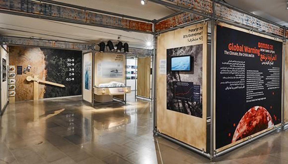
The exhibition, which the museum has been working on for over a year, guides the visitors to the sea, land, glaciers and forests, in the past, present and future. It presents current scientific findings and basic concepts in the field in simple terms and through interactive means, such as videos, thermal cameras that expose thermal gases that surround us, and more.

It uncovers the dire consequences of the climate crisis here in Israel and worldwide, and illustrates the impact of our daily choices as individuals. Everything is not lost; the exhibition illuminates how we can counteract the changes that are causing the crisis and reduce the harm caused to us and the environment.

The various stations of the exhibition show how popular tourist spots may look like in 20 years from now, what the atmospheric composition was thousands of years ago, and what it may be in a few decades from now. The connection between allergies and global warming is explained, as well as what it will be like when sea level reaches our shoulders. You can even check what your personal carbon footprint is, by the help of an online calculator which was developed especially for the exhibition and is the first of its kind in Israel.


What do greenhouse gases look like? Judi Lax explains big concepts in simple language.

Fostering Change Agents

The new exhibition does not, however, intend to scare us into passivity: “We wish to increase the awareness surrounding our daily choices, such as what to eat, how to travel, what to buy and what not to buy. These things have implications and a price beyond the cost of the purchase itself. Oftentimes, people hear about the climate crisis and say, “Ok, but how does this relate to me?” We wish to impart that, although a lot [of damage] has accumulated, it is not all lost. We also have a hand in the matter, and can undo some of the damage,” explains Hadas Zemer Ben-Ari, the exhibition’s curator and designer. “Along with the experience of visiting the exhibition, we strive to make our visitors agents of change, who will spread the message outside the walls of the museum and inspire many others to work for the change that we so desperately need,” says Prof. Tamar Dayan, Museum Chair at the Steinhardt Museum of Nature.

The Museum joins some of the worlds’ largest museums in the common mission to carry out their social role in educating the wider audience on the topic of the climate crisis and the discussion of the biodiversity crisis and its impacts. Museum Director Alon Sapan explains that museums are capable of illustrating and simulating a complex reality and the processes that led to it, along with predictions for the future, while ensuring the visitors’ experience and encouraging their curiosity. “I hope that the exhibition will inspire questions, and also enlighten individuals on how they contribute to positive change by adjusting their personal habits,” concludes Sapan.


An invitation to change a habit (or three!) at the “Global Warning” exhibition.

Featured image: The exhibition “Global Warning: The Climate, the Crisis and Us” (Photo: Dor Kimchi)

Read more and purchase tickets here >> 

Victoria

Tok Corporate Centre, Level 1,
459 Toorak Road, Toorak VIC 3142
Phone: +61 3 9296 2065
Email: [email protected]

New South Wales

Level 22, Westfield Tower 2, 101 Grafton Street, Bondi Junction NSW 2022
Phone: +61 418 465 556
Email: [email protected]

Western Australia

P O Box 36, Claremont,
WA  6010
Phone: :+61 411 223 550
Email: [email protected]