Author: Hilary

Can AI Help Doctors Make Better Diagnoses?

A new TAU study explores how accurate AI can be when assisting with diagnoses in virtual urgent care.

A new study led by Prof. Dan Zeltzer, a digital health expert from the Berglas School of Economics at Tel Aviv University, compared the quality of diagnostic and treatment recommendations made by artificial intelligence (AI) and physicians at Cedars-Sinai Connect, a virtual urgent care clinic in Los Angeles, operated in collaboration with Israeli startup K Health. The paper was published in Annals of Internal Medicine and presented at the annual conference of the American College of Physicians (ACP). This work was supported by funding from K Health.

AI vs. Physicians in Virtual Care

Prof. Zeltzer explains: “Cedars-Sinai operates a virtual urgent care clinic offering telemedical consultations with physicians specializing in family and emergency care. Recently, an AI system was integrated into the clinic—an algorithm based on machine learning that conducts initial intake through a dedicated chat incorporates data from the patient’s medical record and provides the attending physician with detailed diagnostic and treatment suggestions at the start of the visit -including prescriptions, tests, and referrals. After interacting with the algorithm, patients proceed to a video visit with a physician who ultimately determines the diagnosis and treatment. To ensure reliable AI recommendations, the algorithm—trained on medical records from millions of cases—only offers suggestions when its confidence level is high, not recommending about one out of five cases. In this study, we compared the quality of the AI system’s recommendations with the physicians’ actual decisions in the clinic”.


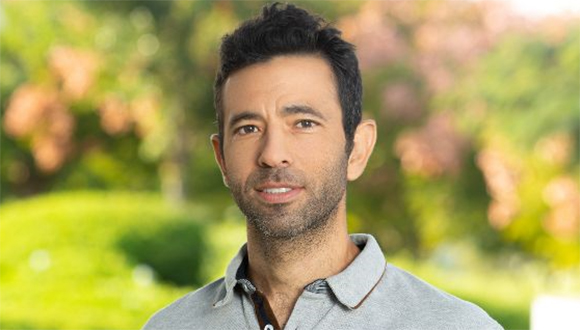
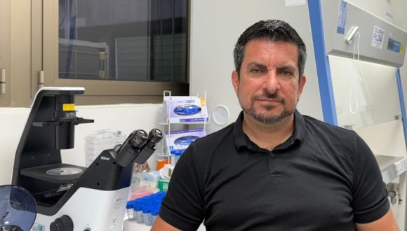
Prof. Dan Zeltzer (Photo courtesy of Richard Haldis).

The researchers examined a sample of 461 online clinic visits over one month during the summer of 2024. The study focused on adult patients with relatively common symptoms—respiratory, urinary, eye, vaginal and dental. In all visits reviewed, patients were initially assessed by the algorithm, which provided recommendations, and then treated by a physician in a video consultation. Afterward, all recommendations—from both the algorithm and the physicians—were evaluated by a panel of four doctors with at least ten years of clinical experience, who rated each recommendation on a four-point scale: optimal, reasonable, inadequate, or potentially harmful. The evaluators assessed the recommendations based on the patient’s medical history, the information collected during the visit, and transcripts of the video consultations.

AI Proves More Accurate Than Physicians in Study

The compiled ratings led to interesting conclusions: AI recommendations were rated as optimal in 77% of cases, compared to only 67% of the physicians’ decisions; at the other end of the scale, AI recommendations were rated as potentially harmful in a smaller portion of cases than physicians’ decisions (2.8% of AI recommendations versus 4.6% of physicians’ decisions).  In 68% of the cases, the AI and the physician received the same score; in 21% of cases, the algorithm scored higher than the physician; and in 11% of cases, the physician’s decision was considered better.

The explanations provided by the evaluators for the differences in ratings highlight several advantages of the AI system over human physicians: First, the AI more strictly adheres to medical association guidelines—for example, not prescribing antibiotics for a viral infection; second, AI more comprehensively identifies relevant information in the medical record—such as recurrent cases of a similar infection that may influence the appropriate course of treatment; and third, AI more precisely identifies symptoms that could indicate a more serious condition, such as eye pain reported by a contact lens wearer, which could signal an infection. Physicians, on the other hand, are more flexible than the algorithm and have an advantage in assessing the patient’s actual condition. For example, if a COVID-19 patient reports shortness of breath, a doctor may recognize it as relatively mild respiratory congestion, whereas the AI, based solely on the patient’s answers, might refer them unnecessarily to the emergency room.

A Step Closer to Supporting Doctors

Prof. Zeltzer concludes: “In this study, we found that AI, based on a targeted intake process, can provide diagnostic and treatment recommendations that are, in many cases, more accurate than those made by physicians. One limitation of the study is that we do not know which physicians reviewed the AI’s recommendations in the available chart, or to what extent they relied on the recommendations. Thus, the study only measured the accuracy of the algorithm’s recommendations and not their impact on the physicians. The study’s uniqueness lies in the fact that it tested the algorithm in a real-world setting with actual cases, while most studies focus on examples from certification exams or textbooks. The relatively common conditions included in our study represent about two-thirds of the clinic’s case volume, thus the findings can be meaningful for assessing AI’s readiness to serve as a decision-support tool in medical practice. We can envision a near future in which algorithms assist in an increasing portion of medical decisions, bringing certain data to the doctor’s attention, and facilitating faster decisions with fewer human errors. Of course, many questions remain about the best way to implement AI in the diagnostic and treatment process, as well as the optimal integration between human expertise and artificial intelligence in medicine”.

Other authors involved in the study include Zehavi Kugler, MD; Lior Hayat, MD; Tamar Brufman, MD; Ran Ilan Ber, PhD; Keren Leibovich, PhD; Tom Beer, MSc; and Ilan Frank, MSc. Caroline Goldzweig, MD MSHS, and Joshua Pevnick, MD, MSHS.

This Is Why Children Took Part in Creating Prehistoric Cave Art

TAU study suggests that children were seen as mediators between the physical and spiritual worlds.

 A team of Tel Aviv University researchers from prehistoric archaeology has proposed an innovative hypothesis regarding an intriguing question: Why did ancient humans bring their young children to cave painting sites, deep underground – through dark, meandering, hazardous passages? The researchers explain: “Next to many cave paintings, there is clear evidence of children as young as two. So far, most hypotheses have focused on the educational aspect — learning the community’s traditions and customs. However, we believe that children also played a unique cultural role in these caves: Young children were credited with special qualities in the spiritual world, enabling them to communicate with entities from the beyond – which were believed to be accessible from the depths of the cave”.

The study was conducted by Dr. Ella Assaf, Dr. Yafit Kedar, and Prof. Ran Barkai from the Jacob M. Alkow Department of Archaeology and Ancient Near Eastern Cultures at Tel Aviv University. The paper was published in the journal Arts from MDPI.

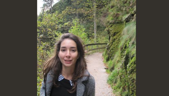
Dr. Ella Assaf.

Children’s Role in Cave Art: More Than Just Education

Dr. Assaf explains: “Cave art created by early humans is a fascinating phenomenon that intrigues many researchers. To date, around 400 caves containing cave art have been discovered, mainly in France and Spain, with the artwork dated between 40,000 and 12,000 years ago. There is solid evidence of children’s participation in the artwork – handprints and finger paintings made by children aged two to twelve. In addition, footprints and handprints of children have been found in some caves, alongside those of adults. This naturally raises the question: Why were the children there? Why were young children taken on exhausting and hazardous journeys deep into the dark, meandering caves with low oxygen levels – crawling through crevices, descending shafts, and climbing rocks to reach their destination?

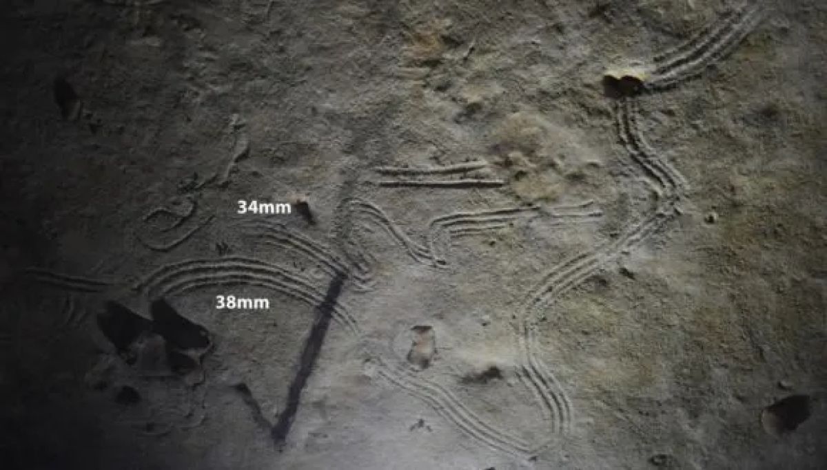
Finger paintings made by children in Rouffignac Cave, 14,000 to 20,000 years ago (Photo courtesy of Dr. Van Gelder).

Dr. Kedar elaborates: “Despite extensive research on cave art, few studies have focused on the presence of children. The prevailing hypothesis is that their participation served an educational purpose – passing down knowledge, traditions, and customs to the next generation. In our study, we argue that children’s involvement had an additional meaning: In fact, they played an important, unique role of their own – direct communication with entities residing in the depths of the earth and otherworldly realms. This study follows our previous works, in which we presented cave artworks as expressions of cosmological approaches, with emphasis on relationships between humans and various entities”.

Children as Mediators Between Worlds in Ancient Rituals

Dr. Assaf adds: “Based on extensive studies about children in indigenous societies, along with new insights into rituals performed in caves with cave paintings, a new understanding is emerging regarding the role of children in the creation of cave art. By integrating data from these research fields, we were able, for the first time, to propose a novel and original explanation for the inclusion of children in creating cave paintings:  The world of childhood differs from that of adults, and children possess a range of unique mental and cognitive traits. For this reason, indigenous cultures worldwide, throughout history and prehistory, have viewed children as ‘active agents’ – mediators between this world and the entities inhabiting the natural world, the underworld, and the cosmos as a whole. In this way, children made a vital contribution to their communities – hunter-gatherers who lived in nature and sought to maintain continuous, respectful relationships with various entities: animals and plants that served as food sources, stones used for toolmaking, ancestral spirits, and more”.

Children’s footprints from Basura Cave, 14,000 years ago (Photo courtesy of Prof. Marco Romano – Romano et al. 2019).

Prof. Barkai: “Many of these societies regarded caves as gateways to the underworld – where, through shamanic rituals, they could communicate with cosmic entities and inhabitants of the underworld, to resolve existential problems. In this context, young children were perceived as liminal beings – belonging to both the realm they had left just recently (before birth) and the world they currently inhabit. Thus, small children were considered particularly suited to bridging the gap between the worlds and delivering messages to non-human entities. In this paper, we connect these insights and propose that children joined adults on journeys into the depths of caves and participated in painting and rituals as part of their role in the community—as ideal mediators with entities from the beyond”.

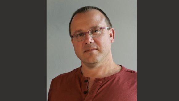
Prof. Ran Barkai.

The Bat’s Walking Cane: How a Tail Becomes a Sensor in the Dark

New research highlights a unique evolutionary adaptation: a bat’s tail acting as a reverse walking cane.

A new study from Tel Aviv University reveals that the Greater Mouse-Tailed Bat (Rhinopoma microphyllum) uses its long tail as a natural tactile sensor to navigate backward in dark caves. The researchers discovered that the bat’s long tail functions as a dynamic sensing tool, enabling it to avoid obstacles and orient itself in complex environments while climbing backward—especially when other sensory abilities like vision and echolocation are limited. The study found that when the tail was numbed with a local anesthetic, the bats’ ability to navigate around obstacles while crawling backward and the speed of their movement decreased significantly.

The research was led by Sahar Hajyahia and Mor Taub, students in the laboratory of Prof. Yossi Yovel of Tel Aviv University’s School of Zoology in the Wise Faculty of Life Sciences and the Sagol School of Neuroscience. The study was published in the scientific journal iScience.

פרופ' יוסי יובל

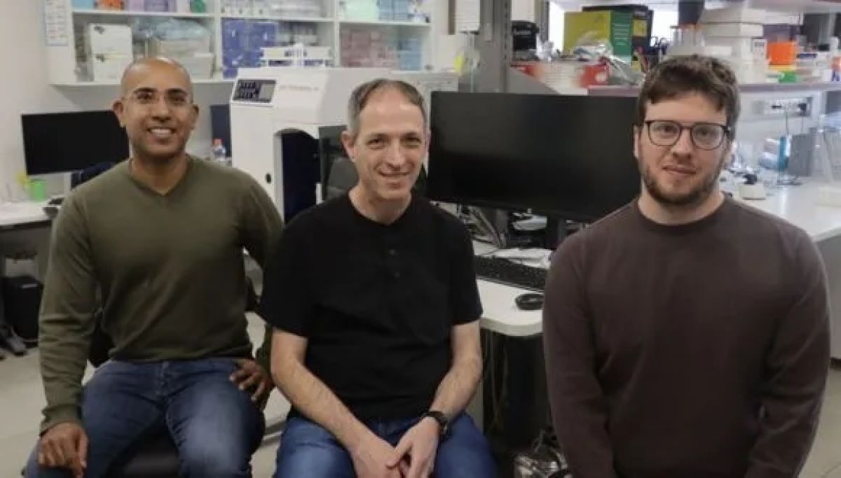
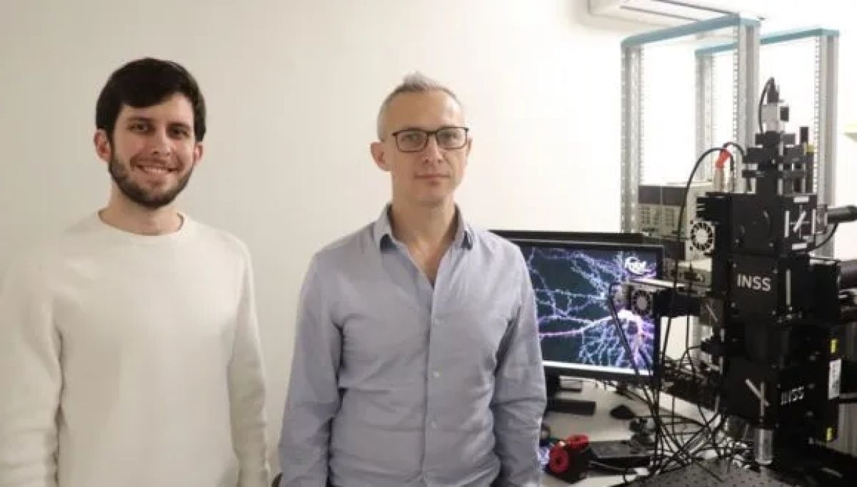
Prof. Yossi Yovel.

Professor Yossi Yovel explains: “During the study, the bats crawled up a vertical maze while moving backward as we recorded their movement using an advanced tracking system. The bats used their tails like a blind walking cane, swinging them from side to side to detect obstacles and climb more safely and efficiently. In contrast, when the tail was numbed, the climbing time increased by an average of 10%, and the bats made more lateral movements instead of moving upward, apparently trying to find their way”.

The researchers also noted that the bats demonstrated a remarkable ability to distinguish between different textures using their tails. They were able to differentiate between a fine wooden grid (1 cm intervals) and a sparser grid (1.5 cm), highlighting the tail’s complex ability to serve as an exceptionally sensitive tactile sensor.

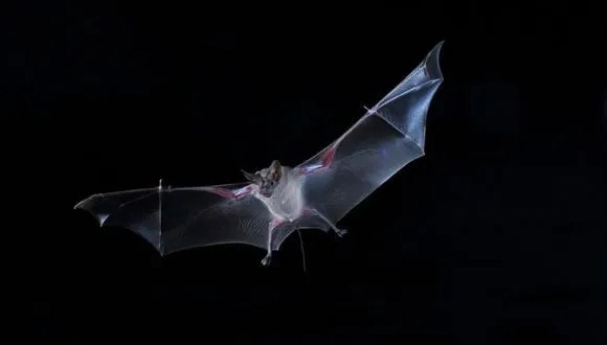
Greater Mouse-Tailed Bat (Photo courtesy of Jens Rydell).

Professor Yossi Yovel concludes: “In most bats, the tail is very short and integrated into the wing membrane. In Rhinopoma, however, the tail remains long and free, and to the best of our knowledge, they are the only bats that use it to sense their immediate surroundings. This is another example of how evolution adapts animals’ senses to meet specific needs—in this case, moving backward in dark places around obstacles and other bats. Many bats crawl backward on dark walls and cannot use their frontal senses like vision and sonar to ‘see’ behind them. One can think of the tail as a sort of reverse sensor for the Rhinopoma. This discovery opens the door to further research on tail usage as a sensor in other animal species. Additionally, the findings could inspire the development of new sensory technologies inspired by nature, such as robotic navigation systems for complex environments”.

Why We Really Exercise: First AI Study Reveals True Motivation

A first-of-its-kind AI study finds 23.9% exercise for appearance and 18.9% for health.

A new study from Tel Aviv University used AI tools for the first time to discover what motivates people to exercise and which strategies are most effective for maintaining physical fitness.

The researchers used tools of artificial intelligence and machine learning to scan thousands of posts on the Reddit social network. They found that 23.9% of the users who engage in sports do so to improve their appearance, 18.9% exercise to maintain their physical health, and 16.9% exercise to maintain their mental health.

The study was led by a team of researchers from TAU’s School of Public Health, Faculty of Medical and Health Sciences : Dr. Michal Shmueli-Scheuer, Yedidya Silverman, Prof. Israel Halperin, and Prof. Yftach Gepner. The paper was published in the Journal of Medical Internet Research (JMIR).

Why Don’t We Exercise More, Even When We Know It’s Crucial?

Prof. Gepner explains: “Researchers in our field usually rely on cumbersome old-school questionnaires, containing inherent biases, to understand why people engage in sports and what strategies help them adhere to physical activity.  It’s an astonishing phenomenon: science tells us that if we put just over two hours a week into physical activity, we can prevent 30% of diseases, improve our quality of life, and extend our lifespan; and yet, less than a quarter of the population actually does this. Why? What have we failed to see?  While we all wish our loved ones good health on their birthday, a wish for ‘good workouts’ is quite rare… But there is a way to be healthy – by exercising. That’s why it’s crucial to understand what really motivates people to engage in physical activity and what helps them stick with it”.

“Our findings are not based on self-reporting, a representative sample, a questionnaire, or a survey. This is, in plain terms, the real reason why people exercise. And the answer is that people mainly exercise to look good. In questionnaires, people claim they want to be healthy, but in reality, they want six-pack abs. These findings are important because they teach us how to address the public, how to persuade people to get off the couch, promote health, and prevent disease”, he adds.

Beyond the question of motivation, the researchers also sought to identify strategies that induce people to engage in physical activity. According to the Reddit posts, 30% rely on workout habits (e.g. morning/evening, every Saturday morning), 13.9% set goals (such as losing weight or running 5 km), 12.1% enjoy the activity itself, 9.7% enjoy socializing during workouts, 8.9% use media (such as YouTube workout videos), 2.8% use fitness apps, and 2.5% have made a financial commitment to adhere to physical activity.

“The results are quite significant”, explains Prof. Gepner. “One strategy is more successful and therefore more recommended than others—creating exercise habits. If you want to be healthier, you need to develop healthy habits, period. Instead of a morning cigarette, drink two glasses of water and go out for a run. 30% is an empirical statistic that is hard to argue with, so as the Head of the Department of Health Promotion, I can confidently say to the public: develop habits and be healthy”.

What Happens Inside a Cell Under Stress?

A novel AI-based method reveals how cells respond to drug treatments.

Researchers from Tel Aviv University have developed an innovative method that can help to understand better how cells behave in changing biological environments, such as those found within a cancerous tumor. The new system, called scNET, combines information on gene expression at the single-cell level with information on gene interactions, enabling the identification of important biological patterns such as responses to drug treatments. The scientific article published in the Nature Methods journal explains how scNET may improve medical research and assist in the development of treatments for diseases. The research was led by PhD student Ron Sheinin under Prof. Asaf Madi, from the Faculty of Medical and Health Sciences and Prof. Roded Sharan, head of the School of Computer Science and AI at Tel Aviv University.

From noisy data to clear insights

Today, advanced sequencing technologies allow the measurement of gene expression at the single-cell level and, for the first time, researchers can investigate the gene expression profiles of different cell populations within a biological sample and discover their effects on the functional behavior of each cell type. One fascinating example is understanding the impact of cancer treatments – not only on the cancer cells themselves but also on the pro-cancer supporting cells or anti-cancer cell populations, such as some cells of the immune system surrounding the tumor.

Despite the amazing resolution, these measurements are characterized by high levels of noise, which makes it difficult to identify precise changes in genetic programs that underlie vital cellular functions. This is where scNET comes into play.

A social network for genes

Ron Sheinin: “scNET integrates single-cell sequencing data with networks that describe possible gene interactions, much like a social network, providing a map of how different genes might influence and interact with each other. scNET enables more accurate identification of existing cell populations in the sample. Thus, it is possible to investigate the common behavior of genes under different conditions and to expose the complex mechanisms that characterize the healthy state or response to treatments”.

Prof. Asaf Madi: “In this research, we focused on a population of T cells, immune cells known for their power to fight cancerous tumors. scNET revealed the effects of treatments on these T cells and how they became more active in their cytotoxic activity against the tumor, something that was not possible to discover before due to the high level of noise in the original data”.

Prof. Roded Sharan: “This is an excellent example of how artificial intelligence tools can help decipher biological and medical data, allowing us to gain new and significant insights. The idea is to provide biomedical researchers with computational tools that will aid in understanding how the body’s cells function, thereby identifying new ways to improve our health”.

In conclusion, scNET demonstrates how combining AI with biomedical research could lead to the development of new therapeutic approaches, reveal hidden mechanisms in diseases, and propose new treatment options.

Closing the Academic Gender Gap in Israel

Some 50 outstanding female TAU graduates have gone abroad for post-docs thanks to a fellowship aiming to increase women’s representation in academia

Though great progress has been made for gender equality in recent decades, academia today still struggles with a severe underrepresentation of women in senior positions. Even at Tel Aviv University, where women make up 58% of PhD students—They compose only 33% of senior faculty. To help enable women to continue in academia, the Presidential Postdoctoral Fellowship for Women is offered to outstanding female TAU graduates to assist them in going abroad for postdoctoral research. Ultimately, many of these brilliant women return to Israel and strengthen the academic landscape through their groundbreaking contributions.

Unique Obstacles

At Tel Aviv University, over 130 talented women complete their doctoral degrees each year. Generally, Israeli researchers who wish to compete for a faculty position at a top Israeli university must go abroad for their postdoctoral research, a requirement which poses particular challenges to women. “Statistics show that the post-doc stage is where the most women fall out of academia,” says Fellowship director Michal Boneh-Mizrahi. “This is true globally, but in Israel women are usually older and already have a family that they must relocate and support, creating further obstacles.”

Aside from social norms that pressure women to prioritize their husbands’ careers, these obstacles are in large part financial. The monetary cost of settling in a new country and fully supporting a family (given that a post-doc’s spouse may not be able to work abroad) is often much higher than any funding given by host institutions which do not take dependents into account. “Funding for women to go abroad to do postdoctoral work should be a high priority,” says TAU Prof. Ines Zucker (Engineering), who received the TAU Fellowship while doing her postdoctoral work on nanomaterials and sustainable water treatment at Yale. “We lose so many talented women from academia because of the financial burden.”   


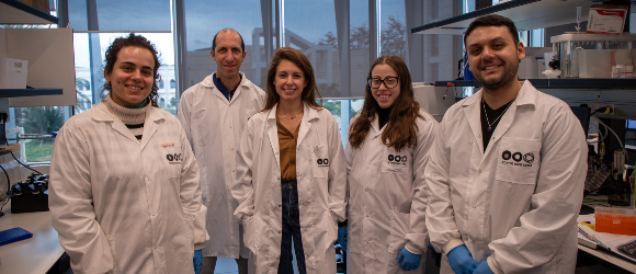
Prof. Zucker with her lab team of student researchers (photo: Rafael Ben-Menashe, TAU).

Providing Support to Advance

Since 2016, Tel Aviv University has enabled 49 outstanding women to conduct postdoctoral research abroad with the Presidential Postdoctoral Fellowship for Women. The two-year Fellowship is offered to 5 female TAU PhD graduates each year. These researchers span a wide range of fields and have traveled to world-renowned institutions including Harvard, Oxford, MIT, Mt. Sinai Hospital and more.

“My time at Harvard was a life-changing experience, both for my career and my family,” says Fellowship recipient Dr. Yifat Naftali Ben Zion (Law), who recently returned to TAU as faculty. “I got to meet so many important people in my field and learn so much that I couldn’t have anywhere else. But it would not have been possible without the Fellowship, as I needed to bring my husband and three young children with me.” 

Dr. Belaynesh Makonen (Education), another former Fellow, spent two years away from her husband and four children at University of Minnesota. “Thanks to the Fellowship, I was able to take time off from my career to accomplish my academic goals.” Dr. Makonen, who was the youngest person ever to become a school inspector in Israel, conducted post-doc research exploring American Jewish identity. She also found a place in the community herself. “I immigrated to Israel from Ethiopia when I was 15; when I got to America, I felt like I was starting over one more time. The Minnesota Jewish community welcomed me with open arms and made sure I had everything I needed.” The support was especially important after Oct. 7 as two of her sons were called to fight in the Gaza war.  

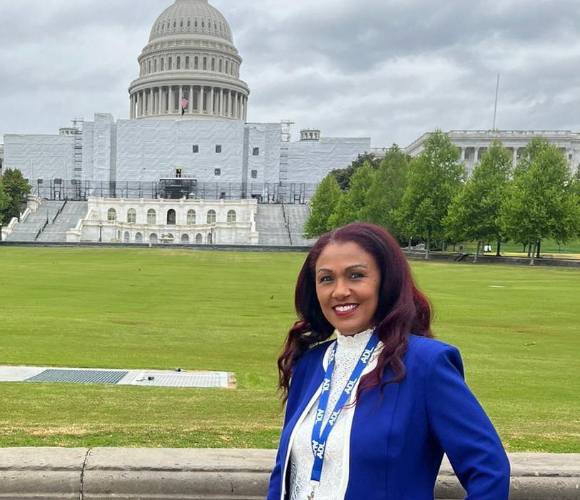
Dr. Belaynesh Makonen traveled to Washington, DC with an Anti-Defamation League leadership program on fighting antisemitism.

Dr. Makonen also took on the responsibility of acting as an advocate and ambassador for Israel during her time abroad. She participated in an ADL leadership program and spoke out about her own identity. “Since I don’t look how Americans expect Israelis to look, I wanted to use my voice to show the diversity of Israel and help spread tolerance within the University of Minnesota.”

At the Forefront of Israeli Research

In addition to acting as ambassadors outside of Israel, the Presidential Fellowship ensures that these women are able to return to Israel and secure top academic positions where their groundbreaking research can have the most impact. Prof. Zucker and Dr. Naftali Ben Zion are two such women who returned to Tel Aviv University from Ivy League universities.

“I had a great time at Yale,” says Prof. Zucker. “I was exposed to the top science in my field and formed lasting professional partnerships.” Since returning to TAU, her lab has become the first in the world to research microplastics and other byproducts of industry in the environment rather than controlled lab conditions; their methods are now used internationally. Her other projects also continue to focus on removal of pollutants from water in affiliation with the TAU Environmental School and the Jan Koum Center for Nanoscience and Nanotechnology.  

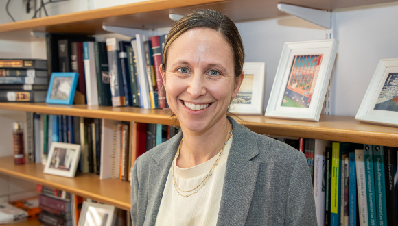
Prof. Naftali Ben Zion researches financial law at Tel Aviv University’s Law Faculty (Photo: Rafael Ben Menashe, TAU).

“I feel very at home here,” says Prof. Zucker, who received all three of her degrees from Tel Aviv University. “I especially appreciate that the schools are set up to encourage collaboration, allowing for incredibly diverse applications of my research.”

“The greatest strength of Israeli universities and TAU specifically is our community,” agrees Prof. Naftali Ben Zion, who researches private law with a specialization in fiduciary law, or the legal framework governing the responsibilities of those entrusted with managing others’ interest. This field of law is relatively unexplored in Israel, and her work has already been cited by the Israeli Supreme Court.

“There’s such a unique amount of support and cooperation here, which creates better academic work. The sharing and mutual development of ideas are the foundation of strong academia.”

Can Stem Cells Restore Bone Marrow?

Doubling adult stem cells for bone marrow and immune system regeneration.

An international research team, led by scientists from Tel Aviv University and Sheba Medical Center, has unveiled an innovative method for activating adult stem cells from human bone marrow, enabling their expansion outside the body for use in bone marrow regeneration and the construction of a new blood and immune system.

The findings, published in the prestigious journal Nature Immunology, represent a breakthrough that could significantly improve transplant success rates for patients who have undergone intensive chemotherapy, suffer from genetic disorders, or require a bone marrow transplant but are unable to source a sufficient number of stem cells from a donor.

The study was led by Dr. Tomer Itkin from the Faculty of Medical and Health Sciences and the Sagol Center for Regenerative Medicine at Tel Aviv University, and the Neufeld Cardiac Research Institute at Sheba Medical Center, Tel Hashomer. The research also included contributions from leading medical institutions worldwide, including Weill Cornell Medical College and Hospital in New York, the Memorial Sloan Kettering Cancer Center (MSKCC), Mount Sinai Hospital, the University of Toronto Medical Center, and the Fred Hutchinson Cancer Research Center in Seattle.

Dr. Tomer Itkin.

Switching On Stem Cells

In the study, which is based on a comprehensive big data analysis of RNA sequencing and epigenetic DNA sequencing, the researchers identified a key protein—the Fli-1 transcription factor—that activates stem cells of the immune and blood system. These stem cells are highly active when sourced from umbilical cord blood but remain in a “dormant” and inactive state when obtained from adult bone marrow donors. Using modified mRNA technology—the same technology used to develop COVID-19 vaccines—the researchers successfully “awakened” the adult stem cells, allowing them to divide in a controlled manner without cancer risk. The activated cells were expanded on endothelial cells, which mimic the blood vessels that support stem cells in the bone marrow environment, demonstrating an enhanced ability to integrate and restore blood production under transplant conditions.

According to Dr. Itkin, This new method significantly expands the available pool of stem cells for transplantation without relying on rare bone marrow donors. Additionally, the method can be used to treat patients whose stem cells have undergone genetic correction, such as those with thalassemia and hereditary anemia, as well as patients who have undergone multiple rounds of chemotherapy and have an insufficient number of stem cells for autologous transplantation“.

The key takeaway from the study is that activating stem cells through molecular programming, rather than arbitrary cell transplantation, substantially improves the success rates of regenerative treatments. The next stage of research involves testing the method in clinical trials to bring this groundbreaking technology into widespread therapeutic use. Furthermore, the researchers plan to apply the same therapeutic approach to regenerate additional tissues, including those without existing adult stem cells, such as the heart.


New PTEN Research Could Transform Autism and Cancer Studies

Is PTEN the key to advancing autism and cancer research?

A novel scientific method developed at Tel Aviv University promises to accelerate our understanding of the gene PTEN, a key player in cellular growth. This breakthrough will help scientists better understand how cells grow and divide, potentially leading to advancements in the treatment of various conditions, including developmental disorders and various forms of cancer.

The study, led by Dr. Tal Laviv in the Faculty of Medical and Health Sciences at Tel Aviv University, was published in the prestigious journal Nature Methods.

The research team explains that cells in the human body constantly adjust their size and rate of division to adapt to their environment throughout life. This process is crucial for normal development, as cells go through periods of precise growth regulation. When this process is disrupted, it can lead to severe diseases such as cancer and developmental disorders.

In the brain, regulating cellular growth is especially critical during early brain development, which occurs in the first years of life. Many genes are involved in this regulation, but one gene in particular—PTEN (Phosphatase and Tensin Homologue)—plays a central role. Mutations in PTEN are linked to a variety of conditions, including autism, epilepsy, and cancer.

PTEN’s Impact Explained

Dr. Tal Laviv explains: “Many studies have shown that PTEN is essential for regulating cell growth in the brain by providing a stop signal. This means PTEN activity is crucial for maintaining cells at their proper size and state. There is growing evidence that mutations in PTEN, which reduce its activity, contribute to diseases like autism, macrocephaly, cancer, and epilepsy. Despite the critical role PTEN plays in cellular function, scientists have had limited tools to measure its activity. For example, it wasn’t to directly measure PTEN activity in an intact brain, which would greatly help our understanding of its role in health and disease”.

Dr. Laviv and his research team, led by MD-PhD student Tomer Kagan, have developed an innovative tool that directly measures PTEN activity with high sensitivity in various research models, including in the intact brains of mice. This groundbreaking technology, which combines advancements in genetic tools and microscopy, will allow scientists to gain deeper insights into why PTEN is so crucial for normal brain development. It could also improve our understanding of how PTEN-related diseases, such as cancer and autism, develop.

The researchers predict that this new tool will enable the development of personalized therapeutics by monitoring PTEN activity in various biological settings. Additionally, it could help identify diseases at earlier stages, potentially leading to faster and more effective treatments.

Gender-Specific Damage in Rare Genetic Syndrome

.ADNP Protein Causes Different Brain Damage in Males and Females.

Researchers at Tel Aviv University, led by Prof. Illana Gozes, examined the effects of different mutations in the ADNP protein, which is essential for normal brain development and aging, on the brain cells of mice — distinguishing between males and females. To their surprise, they found that the defective protein affects completely different mechanisms in the two sexes: in males, the damage occurs in a mechanism that protects the structure of proteins, which in turn disrupts the process of neurogenesis — the production of new brain cells from stem cells — a process crucial for memory and learning. In females, on the other hand, the mechanism that regulates energy within the cell is impaired, preventing the brain from receiving sufficient energy. All of these processes are essential for maintaining memory and learning functions, and their disruption causes significant impairment in both sexes, leading to the development of incurable brain diseases such as Alzheimer’s, in which ADNP is also found to be defective.

The research was conducted by Prof. Illana Gozes, Dr. Gidon Karmon, and doctoral student Guy Shapira from the and the  at Tel Aviv University. Additional contributors to the study include Prof. Noam Shomron, Dr. Gal Hacohen-Kleiman, doctoral student Maram Ganaiem from the Faculty of Medical and Health Sciences, Dr. Shula Shazman from the Department of Mathematics and Computer Science at the Open University, and researchers from the University Hospital of Thessaloniki in Greece. The study was published in the prestigious journal Molecular Psychiatry from Nature.

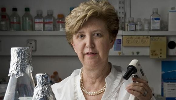
Prof. Illana Gozes.

Prof. Gozes stated: “The ADNP protein was discovered in my lab, and we have been researching it for many years. We found that it is critical for brain development and plays a protective role in neurodegenerative diseases like Alzheimer’s. Additionally, it was found that defects in the ADNP gene cause ADNP syndrome, a rare genetic disorder associated with developmental delays, learning disabilities, and symptoms of autism. In parallel, we are developing the experimental drug Davunetide, which is based on a fragment of the ADNP protein. In this study, we aimed to examine whether ADNP is involved in the process known as ‘neurogenesis’ — the formation of new neurons from stem cells in the adult brain, a process essential for memory and learning. We focused on the hippocampus, a brain region crucial for memory, in adult mice”.

Using genetic engineering, the researchers established two mouse models reflecting different forms of ADNP syndrome: mice that express only half the normal amount of ADNP, with only one active allele in the DNA instead of two, which are typically inherited from both parents and mice with a mutation in the ADNP gene that truncates the protein production process, resulting in a shorter-than-normal ADNP protein.

The researchers note that the most severely affected children with ADNP syndrome are those with the mutation that produces the truncated protein. Additionally, neurogenesis was examined in a control group of healthy mice.

To track the course of neurogenesis, a substance was injected into the mice, staining the DNA of brain cells participating in the process. The data were analyzed using computational bioinformatics methods, proving that ADNP plays a crucial role in neurogenesis. Furthermore, a significant difference was found between how ADNP functions in males versus females. First, in healthy mice, neurogenesis was more active in males than females, while in male mice with an ADNP mutation, neurogenesis was reduced to the same level as in females. A fundamental difference between the sexes was also identified in an additional research method: RNA sequencing of all genes in the hippocampus of mice with the truncated ADNP protein.

How ADNP Protein Breaks Brains by Gender

Prof. Gozes explains: “There was almost no overlap. The damage to the ADNP protein affected completely different mechanisms in male and female brains. The explanation for this phenomenon is that in males, one of the functions of ADNP is to regulate a mechanism that maintains protein structure (unfolded protein response), which in turn regulates neurogenesis. The ADNP gene is a master regulator of this entire mechanism in male brains, and when it is defective, the process is significantly impaired. In females, however, the ADNP protein enters the mitochondria — the cell’s energy powerhouse — and when the mutation alters the protein’s structure, less ADNP can enter the mitochondria. As a result, energy production in the cell is likely impaired, disrupting brain function, which requires a large amount of energy”.

As part of the study, the researchers also tested the effectiveness of the experimental drug Davunetide, based on the NAP fragment of the ADNP protein, in treating affected mice. They observed a positive effect in all cases, with particularly significant neurogenesis recovery in the model where mice had only half the normal ADNP levels.

Promising Drug for ADNP and Beyond

Prof. Gozes concludes: “Our research shows that ADNP is closely linked to neurogenesis and that it functions differently in males and females — a finding that has also emerged in previous studies. Additionally, we found that Davunetide, the drug that we discovered and are developing, is effective. We aim to soon begin a clinical trial in children with ADNP syndrome (ADNP deficiency). We hope that in the future, the drug will also help Alzheimer’s patients — in whom we previously found sex-based differences — as well as other neurodegenerative diseases where ADNP is impaired. Notably, the rare and incurable disease Progressive Supranuclear Palsy (PSP), which has pathological similarities to Alzheimer’s disease, showed significant improvement in women treated with Davunetide in our previous clinical study”.

The pharmaceutical development is being carried out by ExoNavis Therapeutics Ltd under a licensing agreement with Ramot, Tel Aviv University’s technology transfer company. Prof. Gozes serves as Vice President for Drug Development at the company.

TAU Breaks Israeli Record in Space

Tevel 2: Israel’s Largest Satellite Constellation Launch.

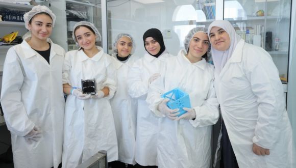
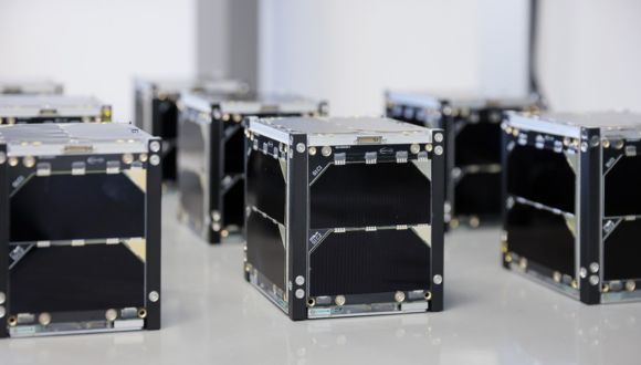
The Ministry of Innovation, Science, and Technology, Tel Aviv University, and nine municipalities across Israel have announced an unprecedented milestone in space research and scientific education: a constellation of nine research nanosatellites—the largest Israeli satellite constellation ever—has been launched into space from Vandenberg Space Force Base in California. The nanosatellites, each measuring 10×10×11.3 cm, were designed and built by high school students from across the country. They were launched into low Earth orbit at an altitude of 500 kilometers aboard a SpaceX Falcon 9 rocket.

Gila Gamliel, Minister of Innovation, Science, and Technology: “The Tevel 2 project symbolizes the pinnacle of Israeli innovation, combined with scientific and technological education. This is an especially emotional moment for me — to see young people from all sectors of Israeli society working together, transforming a dream into reality, and making history in space. What these talented students have accomplished is nothing short of amazing — they have built the largest Israeli satellite constellation ever launched! I am especially proud of the first-ever Druze satellite and the moving memorial mission of the Sha’ar HaNegev satellite. Our substantial investment in this initiative is not just an investment in technology — it is an investment in you, the next generation of Israeli space scientists and engineers”.

Tevel2 students testing environmental conditions at the satellite laboratory at Tel Aviv University (photo credit: Ronen Horesh).

Scientific and Educational Innovation

The revolutionary Tevel 2 project combines scientific innovation with a pioneering educational and social vision. This 10.5m NIS project — initiated and funded by the Israel Space Agency at the Ministry of Innovation, Science, and Technology — connects different communities within Israeli society and promotes equal opportunities in science and technology education, while combining scientific innovation with educational and social objectives.

The satellites were built under the guidance of Tel Aviv University’s Faculty of Engineering, in R&D centers established in nine municipalities throughout the country, uniquely incorporating diverse populations: five Jewish municipalities (Yeruham, Sha’ar HaNegev, Ma’ale Adumim, Givat Shmuel, and Herzliya), three Arab municipalities (Ein Mahil, Tayibe, and Kafr Qara) and one Druze municipality (Yarka).

A representative photo of students in the Tevel2 project. The project includes Jewish, Arab, and Druze students from 9 municipalities across the country (photo creditSivan Shachor).

Brigadier General (Res.) Uri Oron, Director General of the Israel Space Agency: “The Tevel 2 project is a testament to the power of Israeli innovation. We see here a winning combination of scientific excellence, technological education, and societal integration. These satellites will not only provide crucial scientific data; they will also serve as an inspiration for the next generation of Israeli space scientists”.

Prof. Meir Ariel.

Groundbreaking Scientific Research

The satellites, built in special clean rooms in nine municipalities across the country, will perform an important scientific mission.

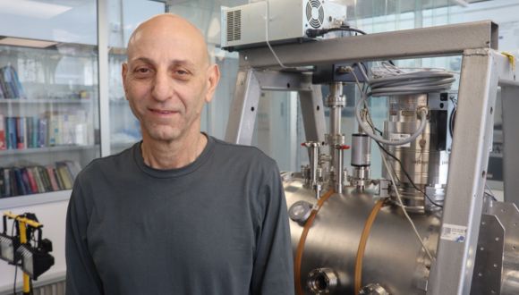
Prof. Meir Ariel, Head of the Space Engineering Center at Tel Aviv University’s Faculty of Engineering, explains: “The satellite constellation is designed to measure cosmic radiation in orbit. Cosmic radiation consists of high-energy particles (mainly protons) originating from the Sun, and events in the deep universe, such as supernova explosions. While life on Earth is relatively protected from this radiation by the planet’s magnetic field and atmosphere, astronauts in space and electronic systems on satellites are exposed to this potentially harmful radiation”.

Tevel2 satellites are ready to be sent to the US for integration into SpaceX Falcon 9 launcher after undergoing environmental tests at the Satellite Laboratory at Tel Aviv University (photo credit: Ronen Horesh).

The nanosatellites, launched as a clustered swarm from the Space Force base in California, are expected, over time, to disperse randomly in their orbit, ultimately creating an accurate high-resolution map of cosmic radiation at every point above Earth’s surface. Another experiment will examine the radiation’s effects on the satellites’ electronic components. The data will be analyzed by the students under the guidance of researchers from Tel Aviv University and the Soreq Nuclear Research Center, where the space radiation sensors were developed.

It should be noted that a significant part of this project is dedicated to promoting science education and strengthening Israel’s social periphery. The satellites were built by outstanding high school students who joined the program in the 10th grade and are now completing the 12th grade. The students worked in R&D centers, complete with clean rooms, established by Tel Aviv University in the nine participating municipalities specified above. Satellite communication stations connected to the main station at Tel Aviv University were set up in Herzliya, Yeruham, Givat Shmuel, and Sha’ar HaNegev, enabling students to control and receive data from the satellites.

One of the program’s main goals is to promote scientific education in Israel’s periphery and provide students from remote regions with an equal opportunity to participate and excel in a national project. This is a key aspect of the visions of the Ministry of Innovation, Science, and Technology, the Israel Space Agency, and Tel Aviv University – all deeply committed to Israeli society and working to reduce social inequality and provide equal opportunities through a variety of affirmative action programs and extensive community initiatives.

Students work on the satellite in the clean room (photo credit: Tapuach Pais, Tayibe).

Commemoration and Remembrance

In addition to its scientific tasks, the project also includes a unique memorial mission: Sha’ar HaNegev’s satellite will telemetrically transmit the names of all Israeli civilians and soldiers killed from October 7, 2023, through December 2024, with the signal displayed continuously, throughout the satellite’s lifespan, on the website of the Israel Space Agency. This unique commemoration holds special significance, since the Sha’ar HaNegev Regional Council suffered great losses in the October 7 attack, with its Mayor Ophir Libstein ז”ל killed by terrorists while defending his community. Moreover, the Sha’ar HaNegev students who participated in the project itself had been evacuated from their homes for many months.

The satellites will remain in orbit for approximately three years, during which they will provide essential scientific data and serve as an inspiration for the next generation of Israeli space scientists and engineers.

Victoria

Phone: +61 3 9296 2065
Email: [email protected]

New South Wales

Phone: +61 02 7241 8711
Email: [email protected]

Western Australia

Phone: :+61 411 223 550
Email: [email protected]