Tag: Biology

TAU Discovery Decodes a Rare Neurological Disease

This breakthrough could pave the way for neurological treatments.

Researchers at Tel Aviv University have developed an innovative research model that allowed them to decode the mechanism underlying a severe and rare neurological disease. The disease is characterized by symptoms such as epilepsy, developmental delay, and intellectual disability.

According to the researchers: “Decoding the disease mechanism is a critical step toward developing treatments targeting specific cellular functions for this disease and other conditions with similar mechanisms affecting cellular energy production”.

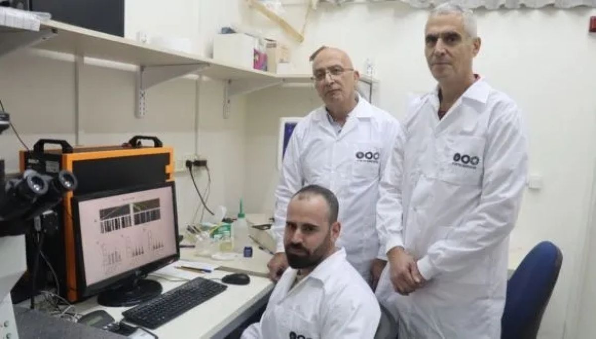
The research was led by Tel Aviv University’s Prof. Abdussalam Azem, Dean of the Wise Faculty of Life Sciences, in collaboration with Prof. Uri Ashery and PhD student Eyal Paz from the School of Neurobiology, Biochemistry and Biophysics at the Wise Faculty of Life Sciences and the Sagol School of Neuroscience. Additional contributors included Dr. Sahil Jain and Dr. Irit Gottfried from the School of Neurobiology, Biochemistry, and Biophysics at Tel Aviv University, Dr. Orna Staretz-Chacham from the Faculty of Health Sciences at Ben-Gurion University, Dr. Muhammad Mahajnah from the Technion, and researchers from Emory University in Atlanta, USA. The findings were published in the prominent journal eLife.

TIMM50 Mutation Linked to Rare Brain Disorders

Prof. Azem explains: “The disease we studied is caused by a mutation in a protein called TIMM50, which plays a crucial role in importing other proteins into the mitochondria—the organelle considered the cell’s energy powerhouse. The human mitochondria operate with about 1,500 proteins (approximately 10% of all human proteins), but only about 13 of them are produced within the mitochondria itself. The rest are imported externally through various mechanisms. In recent years, mutations in the TIMM50 protein, which is responsible for importing about 800 proteins into the mitochondria, were found to cause severe and rare neurological disease with symptoms like epilepsy, developmental delay, and intellectual disability”.

Prof. Ashery adds: “Protein import into the mitochondria has been extensively studied over the years, but how a mutation in TIMM50 affects brain cells was never tested before. To investigate this for the first time, we created an innovative model using mouse neurons that mimics the disease caused by the TIMM50 protein mutation. In this study, we significantly reduced the expression of the protein in mouse brain cells and observed its impact on the cells”.

How Does a Protein Defect Link Energy Loss to Epilepsy?

Eyal Paz explains: “The impairment of the protein led to two main findings: a reduction in energy production in the neurons, which could explain the developmental issues seen in the disease and an increase in the frequency of action potentials (the electrical signals that transmit information along neurons and enable communication between them). This increase in action potential frequency is known to be associated with epilepsy. The change in frequency is likely caused by significant damage to two proteins that function as potassium channels. Imbalances in potassium levels can lead to life-threatening conditions, such as arrhythmias, cardiac arrest, and muscle weakness, potentially leading to paralysis. These potassium channels may serve as potential targets for future drug treatments for the disease”.

Prof. Azem concludes: “Our study decodes the mechanism of a severe and rare neurological disease caused by a mutation in a protein critical for importing proteins into the mitochondria. Understanding the mechanism is a crucial step toward treatment, as it enables the development of drugs targeting the specific issues identified. Additionally, we created a new research model based on mouse neurons that significantly advances the study of protein import into mitochondria in brain cells. We believe that our findings, combined with the innovative model, will enable more in-depth research and the development of treatments for various neurological diseases caused by similar mitochondrial dysfunction mechanisms”.

How Does the Brain Keep Calm?

New Insight into Brain Stability: The Key Role of NMDA Receptors

Researchers at Tel Aviv University have made a fundamental discovery: the NMDA receptor (NMDAR)—long studied primarily for its role in learning and memory—also plays a crucial role in stabilizing brain activity. By setting the “baseline” level for activity in neural networks, the NMDAR helps maintain stable brain function amidst continuous environmental and physiological changes. This discovery may lead to innovative treatments for diseases linked to disrupted neural stability, such as depression, Alzheimer’s disease, and epilepsy.

The study was led by Dr. Antonella Ruggiero, Leore Heim, and Dr. Lee Susman from Prof. Inna Slutsky’s lab at the Faculty of Medical and Health Sciences at Tel Aviv University. Prof. Slutsky, who is also affiliated with the Sagol School of Neuroscience, heads the Israeli Society for Neuroscience and directs the Sieratzki Institute for Advances in Neuroscience. Additional researchers included Dr. Ilana Shapira, Dima Hreaky, and Maxim Katsenelson from the Faculty of Medical and Health Sciences at Tel Aviv University, and Prof. Kobi Rosenblum from the University of Haifa. The study was published in the prestigious journal Neuron.

“In recent decades, brain research has mainly focused on processes that allow information encoding, memory, and learning, based on changes in synaptic connections between nerve cells”, says Prof. Slutsky.

“But the brain’s fundamental stability, or homeostasis, is essential to support these processes. In our lab, we explore the mechanisms that maintain this stability, and in this study, we focused on the NMDAR—a receptor known to play a role in learning and memory”, Slutsky continues.

This comprehensive project used three primary research methods: electrophysiological recordings from neurons in both cultured cells (in vitro) and living, behaving mice (in vivo) within the hippocampus, combined with computational modeling (in silico). Each approach provided unique insights into how NMDARs contribute to stability in neural networks.

Dr. Antonella Ruggiero studied NMDAR function in cultured neurons using an innovative technique called “dual perturbation”, developed in Prof. Slutsky’s lab. “First, I exposed neurons to ketamine, a known NMDAR blocker”, she explains. “Typically, neuronal networks recover on their own after disruptions, with activity levels gradually returning to baseline due to active compensatory mechanisms. But when the NMDAR was blocked, activity levels stayed low and didn’t recover. Then, with the NMDAR still blocked, I introduced a second perturbation by blocking another receptor. This time, the activity dropped and recovered as expected, but to a new, lower baseline set by ketamine, not the original level”. This finding reveals the NMDAR as a critical factor in setting and maintaining the activity baseline in neuronal networks. It suggests that NMDAR blockers may impact behavior not only through synaptic plasticity but also by altering homeostatic set points.

Building on this discovery, Dr. Ruggiero sought to uncover the molecular mechanisms behind the NMDAR’s role in tuning the set point. She identified that NMDAR activity enables calcium ions to activate a signaling pathway called eEF2K-BDNF, previously linked to ketamine’s antidepressant effects.

How NMDARs Set the Brain’s Activity Baseline

Leore Heim investigated whether the NMDAR similarly affects baseline activity in the hippocampus of living animals. A major technical challenge was administering an NMDAR blocker directly to the hippocampus without affecting other brain areas, while recording long-term activity at the individual neuron level. “Previous studies often used injections that delivered NMDAR blockers across the entire brain, leading to variable and sometimes contradictory findings,” he explains. “To address this, I developed a method combining direct drug infusion into the hippocampus with long-term neural activity recording in the same region. This technique revealed a consistent decrease in hippocampal activity across states like wakefulness and sleep, with no compensatory recovery as seen with other drugs. This strongly supports that NMDARs set the activity baseline in hippocampal networks in living animals”.

Mathematician Dr. Lee Susman created computational models to answer a longstanding question: Is brain stability maintained at the level of the entire neural network, or does each neuron individually stabilize itself? “Based on the data from Antonella and Leore’s experiments, I found that stability is maintained at the network level, not within single neurons,” he explains. “Using models of neural networks, I showed that averaging activity across many neurons provides computational benefits, including noise reduction and enhanced signal propagation. However, we need to better understand the functional significance of single-neuron drift in future studies”.

Prof. Slutsky adds: “We know that ketamine blocks NMDARs, and in 2008, it was FDA-approved as a rapid-acting treatment for depression. Unlike typical antidepressants like Cipralex and Prozac, ketamine acts immediately by blocking NMDARs. However, until now, it wasn’t fully understood how the drug produced its antidepressant effects. Our findings suggest that ketamine’s actions may stem from this newly discovered role of NMDAR: reducing the activity baseline in overactive brain regions seen in depression, like the lateral habenula, without interfering with homeostatic processes. This discovery could reshape our understanding of depression and pave the way for developing innovative treatments”.

Hyperbaric Oxygen Therapy: A Promising Treatment for PTSD Symptoms

Biological damage in PTSD sufferers can be treated with a specialized protocol.

Researchers at Tel Aviv University and the Sagol Center for Hyperbaric Medicine and Research at the Shamir Medical Center have demonstrated that hyperbaric oxygen therapy (HBOT) improves the condition of PTSD sufferers who have not responded to psychotherapy or psychiatric medications. The researchers: “Our unique therapeutic protocol affects the biological brain ‘wound’ associated with PTSD, and effectively reduces typical symptoms such as flashbacks, hypervigilance, and irritability. We believe that our findings give new hope to millions of PTSD sufferers and their families, all over the world”.

The study was led by Prof. Shai Efrati and Dr. Keren Doenyas-Barak from the Faculty of Medical and Health Sciences at Tel Aviv University and the Sagol Center for Hyperbaric Medicine and Research at the Shamir Medical Center. Other contributors include Dr. Ilan Kutz, Gabriela Levi, Dr. Erez Lang, Dr. Amir Asulin, Dr. Amir Hadanny, and Dr. Ilia Beberashvili from the Shamir Medical Center, and Dr. Kristoffer Aberg and Dr. Avi Mayo from the Weizmann Institute. The paper was published in The Journal of Clinical Psychiatry.

“At present, we treat hundreds of PTSD sufferers every day”

Prof. Efrati: “Due to our unfortunate circumstances, Israel has become a global leader in the field of PTSD. Before the Hamas attack on Oct. 7, 2023, approximately 6,000 IDF veterans had been recognized as PTSD sufferers, with many others, both soldiers and citizens, not yet acknowledged by the authorities. Following Oct. 7 and the ensuing war, these numbers have risen sharply. Tens of thousands of soldiers, and much larger numbers of civilians, are likely to be diagnosed with PTSD. The world-leading Sagol Center for Hyperbaric Medicine, the largest of its kind in the world, is rising to the challenge – with a comprehensive therapeutic array comprising hyperbaric facilities combined with diverse mental health professionals, psychologists and psychiatrists. At present, we treat hundreds of PTSD sufferers every day, aiming to reach one thousand patients per year”.

Dr. Doenyas-Barak: “PTSD (Post-Traumatic Stress Disorder) is defined as the mental outcome of exposure to a life-threatening event. About 20% of those who have undergone such an experience will develop PTSD, which can lead to substantial social, behavioral, and occupational dysfunctions. In extreme cases, the disorder can severely impact their quality of life, family life, and professional performance. Symptoms include a range of emotional and cognitive changes, nightmares and flashbacks, hypervigilance, irritability, and avoidance – so as not to trigger traumatic experiences. In many cases, PTSD is resistant to psychotherapy and common psychiatric medications. Past studies on therapy-resistant sufferers have found changes in the structure and function of brain tissues, or a ‘biological wound’ that explains such treatment resistance. In our study, we wanted to determine whether hyperbaric therapy can help these patients”.

Testing HBOT for PTSD Relief

The study, which began in 2019 and ended in the summer of 2023, included 98 male IDF veterans diagnosed with combat-associated PTSD, who had not responded to either psychotherapy or psychiatric medications. Participants were divided into two groups: one group received HBOT treatment, breathing pure high-pressure oxygen, while the other underwent the same procedure, but received a placebo treatment, breathing regular air. 28 members of each group completed the process and the following evaluation.

Dr. Doenyas-Barak: “The HBOT was administered in accordance with a unique treatment protocol developed at our Center. Every patient is given a series of 60 two-hour treatments in our hyperbaric chamber, during which they are exposed to pure 100% oxygen at a pressure of 2 atmospheres (twice the normal air pressure at sea level). Our protocol specifies alternately breathing oxygen and regular air: every 20 minutes the patient removes the oxygen mask and breathes regular air for five minutes. The drop in oxygen level, at the tissue level, activates healing processes and thus enhances the therapeutic effect”.

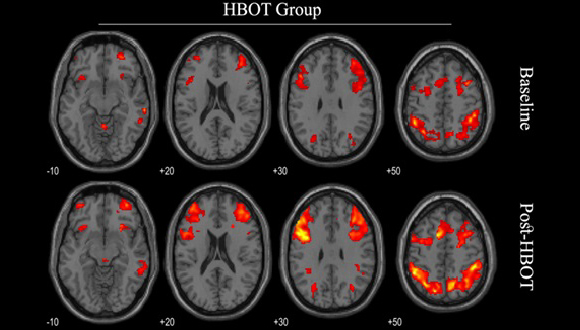
Functional MRI before and after HBOT  Photo credit: The Shamir Medical Center.

Functional MRI before and after HBOT. Photo credit: The Shamir Medical Center.

The results were encouraging, with improvements observed both at the clinical level and in fMRI imaging.  The group that received hyperbaric therapy showed improved connectivity in brain networks, alongside a decline in all typical PTSD symptoms. In the placebo group, on the other hand, no change was observed in either the brain or clinical symptoms. Prof. Efrati: “Our study demonstrated that HBOT induces biological healing in the brain of PTSD sufferers. Curing the biological wound also impacts clinical symptoms. We believe that HBOT, based on the special protocol we have developed, can bring relief to numerous PTSD sufferers worldwide, allowing them to resume a normative life in their community and family”.

Prof. Efrati emphasizes:

“Patients suffering from PTSD should undergo HBOT only at professional hyperbaric centers, where treatment is delivered by multidisciplinary teams experienced in trauma care. Unsupervised, private hyperbaric chambers are unable to provide a proven, effective protocol. Additionally, patients must receive a thorough professional evaluation to ensure they are suitable for HBOT and to determine what additional support is needed throughout their treatment journey”.

Israel’s Ministry of Defense funds HBOT for veterans who need it.

Buzzed but Never Tipsy: Hornets’ Remarkable Alcohol Tolerance

Oriental hornets are the only animals able to drink unlimited amounts of alcohol.

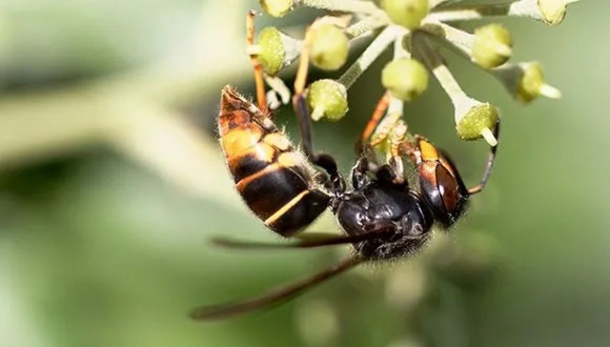
A new study from the School of Zoology  and the Steinhardt Museum of Natural History  at Tel Aviv University has revealed that the Oriental hornet is the only known animal capable of chronically consuming alcohol in high concentrations with almost no negative effects on its health or lifespan. The research team says, “This is a remarkable animal that shows no signs of intoxication or illness even after ingesting huge amounts of alcohol.”

The research was conducted under the leadership of postdoctoral fellow Dr. Sofia Bouchebti from Prof. Eran Levin’s laboratory at Tel Aviv University’s School of Zoology and the Steinhardt Museum of Natural History. The study was published in the journal Proceedings of the National Academy of Sciences of the United States of America (PNAS).

The researchers explain that alcohol is commonly produced in nature through the breakdown of sugars by yeasts and bacteria, primarily found in ripe fruits and nectar. Although alcohol contains nearly twice the amount of energy as sugar, it is toxic to most animals — including us humans — with occasional consumption, and especially with chronic use. Among the animals known to consume alcohol are fruit flies, which show signs of alcohol poisoning even at relatively low concentrations, and treeshrews — mammals native to East Asia that feed on ripe, alcohol-rich fruits — who show symptoms such as fatty liver and other effects indicative of alcoholism after consuming low concentrations of alcohol continuously for several days.

As for humans, many of us like consuming alcohol. Humans domesticated the wine grape around 10,000 years ago, and compared to other animals, we can tolerate and often enjoy consuming relatively high amounts of alcohol. However, as we know, alcohol has significant effects on behavior, cognition, and, of course, health, with a host of diseases linked to its consumption.

Hornets Can Handle Their Liquor

In the new study, the research team tested the Oriental hornet’s ability to consume alcohol and break it down. Dr. Bouchebti explains: “The hornets naturally store yeasts in their digestive system, which provides them with a unique environment that allows the yeast to develop and reproduce, creating new strains. One explanation is that hornets transfer yeasts to fruits, which indirectly contributes to the production of wine. In our study, we labeled the alcohol consumed by the hornets with a heavy carbon isotope. As the alcohol is metabolized, it breaks down into carbon dioxide, which is exhaled. By measuring the amount of labeled carbon dioxide emitted, we were able to estimate the speed at which the alcohol was broken down. The findings were surprising; we were amazed to see the rapid rate at which the hornets metabolized the alcohol”.

In the next stage, the researchers sought to determine whether the Oriental hornet ever becomes intoxicated. Does increased alcohol consumption affect their behavior, for example causing aggression or impacting their nest-building abilities? Here too, the findings were surprising: even when consuming high concentrations of alcohol (80 percent alcohol as the sole source of nutrition) there was no noticeable effect on the hornets’ behavior. In the final phase of the study, the researchers tested whether alcohol had any impact on the hornets’ lifespan and health. Once again, they were amazed to discover that no differences were found between the lifespan of hornets that consumed only alcohol for their entire lives (three months) and hornets that consumed sugar water.

No Hangovers Here

Prof. Levin concludes: “To the best of our knowledge, Oriental hornets are the only animal adapted to consuming alcohol as a metabolic fuel. They show no signs of intoxication or illness, even after chronically consuming huge amounts of alcohol, and they eliminate it from their bodies very quickly. In a bioinformatics analysis of the Oriental hornet’s genome, conducted by Prof. Dorothee Huchon, it was discovered that the hornet possesses several copies of the gene responsible for producing the enzyme that breaks down alcohol; this genetic adaptation may be related to their incredible ability to handle alcohol. We propose that the ancient relationship between hornets and yeast led to the development of this adaptation. Furthermore, while alcohol-related research is highly advanced, with 5.3 percent of deaths in the world linked to alcohol consumption, we believe that, following our research, Oriental hornets could potentially be used to develop new models for studying alcoholism and the metabolism of alcohol”.

Due to Climate Change, More Animals will Become Extinct Outside of Nature Reserves than Within Them

According to int’l study surveying more than 14,000 species of amphibians and reptiles.

A new international study has found that amphibians and reptiles inhabiting the world’s nature reserves, or Protected Areas (PAs), will be better protected against climate change than species found outside of these areas, but are still likely to be harmed.

The research findings provide evidence, on a global scale, of the crucial role Protected Areas play in conserving amphibian and reptile biodiversity under human-induced climate change scenarios. The study reveals that more animals will become extinct because of climate change outside of Protected Areas than inside them — in the world in general and on most individual continents.

Protected Areas as Refuges

Prof. Shai Meiri of Tel Aviv University’s School of Zoology, The George S. Wise Faculty of Life Sciences and The Steinhardt Museum of Natural History took part in the study, in collaboration with leading researchers from 19 countries. The study was published in the prestigious journal Nature Communications.


“Approximately 91% of the amphibian and reptile species we examined are protected, to some degree, in Protected Areas, and this proportion will remain unchanged under future climate change.” – Prof. Shai Meiri


The purpose of the study was to evaluate the effectiveness of existing Protected Areas in protecting the amphibians and reptiles living within them under future climate scenarios, as well as to identify conservation gaps in order to outline a road map for the development of conservation actions based on the current global network of Protected Areas.

“In this study, we collected distribution data for more than 14,000 species of amphibians and reptiles — about 70% of the known species — to perform a global assessment of the conservation effectiveness of Protected Areas in an era of climate change, using species distribution models,” explains Prof. Meiri. “Our analyses revealed that approximately 91% of the amphibian and reptile species we examined are protected, to some degree, in Protected Areas, and that this proportion will remain unchanged under future climate change. Furthermore, species protected in Protected Areas will lose smaller portions of their distribution ranges inside the nature reserve than outside of them. Therefore, the proportion of species within reserves is expected to increase.”

Relative Optimism

However, Prof. Meiri points out, “We predict that more than 300 of the amphibian species and 500 of the reptile species we studied will become extinct due to climate change in the coming decades, and probably also hundreds of species for which we did not have sufficient data to model. Our research highlights the importance of Protected Areas in providing refuge for amphibians and reptiles in face of climate change and points out areas where there are not enough nature reserves that can better preserve biodiversity around the world.”


“Despite the relative optimism emerging from the new research, the models still predict extremely high rates of loss of species and habitats due to climate change. Protected Areas do indeed protect the animals living within them, but nothing is foolproof.” – Prof. Shai Meiri


He adds: “We compiled a comprehensive global database with more than 3.5 million observation records spanning 5,403 amphibian species and 8,993 reptile species from online databases, fieldwork data, museum collections, and published references. For all species in our database, we predicted habitat availability according to current (1960–1990) climate data and future scenarios (for the years 2060–2080) at a high spatial resolution (1 km × 1 km) using species distribution models. Then, we evaluated the effectiveness of Protected Areas in conserving amphibians and reptiles by calculating the coverage of their distribution range inside and outside of Protected Areas, as well as the proportion of species for whom a significant portion of their distribution range (for example, 15% or 30%) is protected in PAs under current and future climate conditions (assuming that the future use of the land remains unchanged over the years — that is, that there will be no conversion of nature reserves into agricultural, industrial, or urban areas.)”

Prof. Meiri concludes: “Our evidence shows that the current global network of Protected Areas already plays an important role in preserving the global biodiversity of amphibians and reptiles, and will continue to do so under the expected future climate. However, many species do not live in the existing Protected Areas. These include, for example, many amphibians and reptiles in Mexico, Jamaica, the Andes, West Africa, South Africa, the southern and northern coast of Turkey, Yemen and other places. Moreover, in our study we could create a model for only about two-thirds of reptile and amphibian species. Good models can’t be created for the rarest species, which are known to be more vulnerable to extinction and less protected in Protected Areas. At the same time, it is important to remember that despite the relative optimism emerging from the new research, the models still predict extremely high rates of loss of species and habitats due to climate change. Protected Areas do indeed protect the animals living within them, but nothing is foolproof.”

Research based on a comprehensive study of 8,000 birds in Israel

Tel Aviv University (TAU) researchers say that climate change may be responsible for changes in the morphology of many birds in Israel over the past 70 years. The body mass of some species decreased while in others body length increased, in both cases increasing the ratio between surface area and volume. The researchers contend that these are strategies to facilitate heat loss to the environment.

“The birds evidently changed in response to the changing climate,” the researchers concluded. “However, this solution may not be fully adequate, especially as temperatures continue to rise.”

The study was led by Professor Shai Meiri and PhD student Shahar Dubiner of the School of Zoology, Wise Faculty of Life Sciences, and the Steinhardt Museum of Natural History at TAU. The paper was published in the scientific journal Global Ecology and Biogeography.

Professor Meiri explains that according to “Bergmann’s rule,” an ecogeographical rule formulated in the 19th century, members of bird and mammal species living in a cold climate tend to be larger than members of the same species living in a warmer climate. This is because the ratio of surface area to volume is higher in smaller animals, permitting more heat loss (an advantage in warm regions), and lower in larger bodies, minimizing heat loss (a benefit in colder climates). Based on this rule, scientists have predicted that global warming will lead to a reduction in animal size, with a possible exception: birds living in the human environment (such as pigeons, house sparrows, and the hooded crow) may gain size due to increased food availability, a phenomenon already witnessed in mammals such as jackals and wolves.

Relying on the vast bird collection preserved by the Steinhardt Museum of Natural History at TAU, the researchers looked for changes in bird morphology over the past 70 years in Israel. They examined approximately 8,000 adult specimens of 106 different species, including migratory birds that annually pass through Israel such as the common chiffchaff, white stork, and black buzzard; resident wild birds like the Eurasian jay, Eurasian eagle-owl, and rock partridge; and commensal birds that live near humans. They built a complex statistical model consisting of various parameters to assess morphological changes — in the birds’ body mass, body length and wing length — during the relevant period.

“Our findings revealed a complicated picture,” Dubiner says. “We identified two different types of morphological changes: some species had become lighter – their mass had decreased while their body length remained unchanged; while others had become longer – their body length had increased, while their mass remained unchanged. These together represent more than half of the species examined, but there was practically no overlap between the two groups – almost none of the birds had become both lighter and longer.

“We think that these are two different strategies for coping with the same problem, namely the rising temperatures. In both cases, the surface area to volume ratio is increased by either increasing the numerator or reducing the denominator, which helps the body lose heat to its environment. The opposite, namely a decrease in this ratio, was not observed in any of the species.”

These findings were observed across the country, regardless of nutrition, and in all types of species. A difference was identified, however, between the two strategies: changes in body length tended to occur more in migrants, while changes in body mass were more typical of non-migratory birds. The very fact that such changes were found in migratory birds coming from Asia, Europe, and Africa suggests that this is a global phenomenon. The study also found that the impact of climate change over time on bird morphology is 10 times greater than the impact of similar differences in temperature between geographical areas.

“Our findings indicate that global warming causes fast and significant changes in bird morphology,” Dubiner concludes. “But what are the implications of these changes? Should we be concerned? Is this a problem, or rather an encouraging ability to adapt to a changing environment? Such morphological changes over a few decades probably do not represent an evolutionary adaptation, but rather certain phenotypic flexibility exhibited by the birds. We are concerned that over such a short period of time, there is a limit to the flexibility or evolutionary potential of these traits, and the birds might run out of effective solutions as temperatures continue to rise.”

Inventive Study to Develop Biological Solutions for Agriculture

TAU and ag-biotech company PlantArcBio to collaborate on development of RNAi-based products.

Genetically improved plants can be a real-life magic stick for solving global famine issues. In a first-of-its-kind study, Ramot, the Technology Transfer Company of Tel Aviv University will cooperate with ag-biotech company PlantArcBio to develop innovative RNAi-based biological solutions for agriculture.

RNAi technology enables a temporary external disruption of RNA (ribonucleic acid) molecules, diminishing the amount of Messenger RNA (mRNA), thus temporarily reducing the expression of specific genes, without modifying or genetically engineering the organism’s DNA. Externally applied RNAi molecules affect specific genes for a specific time period, as required for positive effects like crop protection and yield enhancement. 

Specifically, the research will focus on testing the joint technology’s contribution to the stability of RNAi-based products and their ability to penetrate plants and insects.

Joining Forces

The first-of-its-kind joint study will examine the efficacy of PlantArcBio‘s RNAi technology for agriculture, combined with the unique lipid-based RNA delivery technology developed by Prof. Dan Peer, TAU’s Vice President for R&D, head of the Center for Translational Medicine and a member of both the Shmunis School of Biomedicine and Cancer ResearchGeorge S. Wise Faculty of Life Sciences, and the Center for Nanoscience and Nanotechnology, and a pioneer using RNA to manipulate cells in cancer and other immune related diseases.  


Prof. Dan Peer

“We see great value in contributing to the development of RNAi-based products addressing global issues and providing an ecological and environmentally friendly solution to the global challenges of sustainability in agriculture and food security,” says Peer.

Keren Primor Cohen, CEO of Ramot, believes there is “extensive commercial potential for this combined technology” and welcomes the collaboration with PlantArcBio.

The research will be carried out both at PlantArcBio‘s Laboratories and at Prof. Dan Peer’s Laboratory of Precision NanoMedicine at Tel Aviv University. According to Dror Shalitin, Founder and CEO of PlantArcBio, the results are expected within approximately 12 months.

Tiny insects become “visible” to bats when they swarm

3-D simulations could provide new insights into the evolution of bat echolocation, TAU researchers say

Bats use echolocation to hunt insects, many of which fly in swarms. In this process, bats emit a sound signal that bounces off the target object, revealing its location. Smaller insects like mosquitos are individually hard to detect through echolocation, but a new Tel Aviv University study reveals that they become perceptible when they gather in large swarms. The findings could provide new insights into the evolution of bat echolocation and explain why tiny insects are found in the diets of bats that seem to use sound frequencies that are too high to effectively detect them. The new research was conducted by Dr. Arjan Boonman and Prof. Yossi Yovel at TAU’s Department of Zoology and colleagues at Canada’s Western University. It was published in PLOS Computational Biology on December 12.

Modeling bat vision

Few studies have addressed what swarms of insects — as opposed to single insects — “look” like to bats. To find out, Dr. Boonman and colleagues combined three-dimensional computer simulations of insect swarms with real-world measurements of bat echolocation signals to examine how bats sense swarms that vary in size and density. They found that small insects that are undetectable on their own, such as mosquitos, suddenly become “visible” to bats when they gather in large swarms. They also discovered that the fact that bats use signals with multiple frequencies is well suited to the task of detecting insect swarms. These signals appear to be ideal for detecting an object if more than one target falls inside the echolocation signal beam at once. “Using simulations, we investigated something that could never have been measured in reality,” Dr. Boonman says. “Modeling enabled us to have full control over any aspect of an insect swarm, even the full elimination of the shape of each insect within the swarm.”

From insects to drones

The insect model the researchers used has a tiny mesh (skeleton) and minuscule legs and wings. “We are still adding new features, such as the bat’s acoustic beam or ears, which were not in the original model,” says Prof. Yovel. “We also developed a faster version of the algorithm. All of this will open a new world for us in which we can get echoes even from entire landscapes, so we can learn what a bat or sonar-robot would ‘see’ much more quickly.” The study could also affect technology being developed to improve defense systems. “The algorithms developed for this study could potentially be applied to radar echoes of drone swarms in order to lower the probability of detection by enemy radar,” Dr. Boonman explains. “Since drones are playing an ever more prominent role in warfare, our biological study could spawn new ideas for the defense industry.”

Victoria

Tok Corporate Centre, Level 1,
459 Toorak Road, Toorak VIC 3142
Phone: +61 3 9296 2065
Email: [email protected]

New South Wales

Level 22, Westfield Tower 2, 101 Grafton Street, Bondi Junction NSW 2022
Phone: +61 418 465 556
Email: [email protected]

Western Australia

P O Box 36, Claremont,
WA  6010
Phone: :+61 411 223 550
Email: [email protected]