
Tag: Medicine

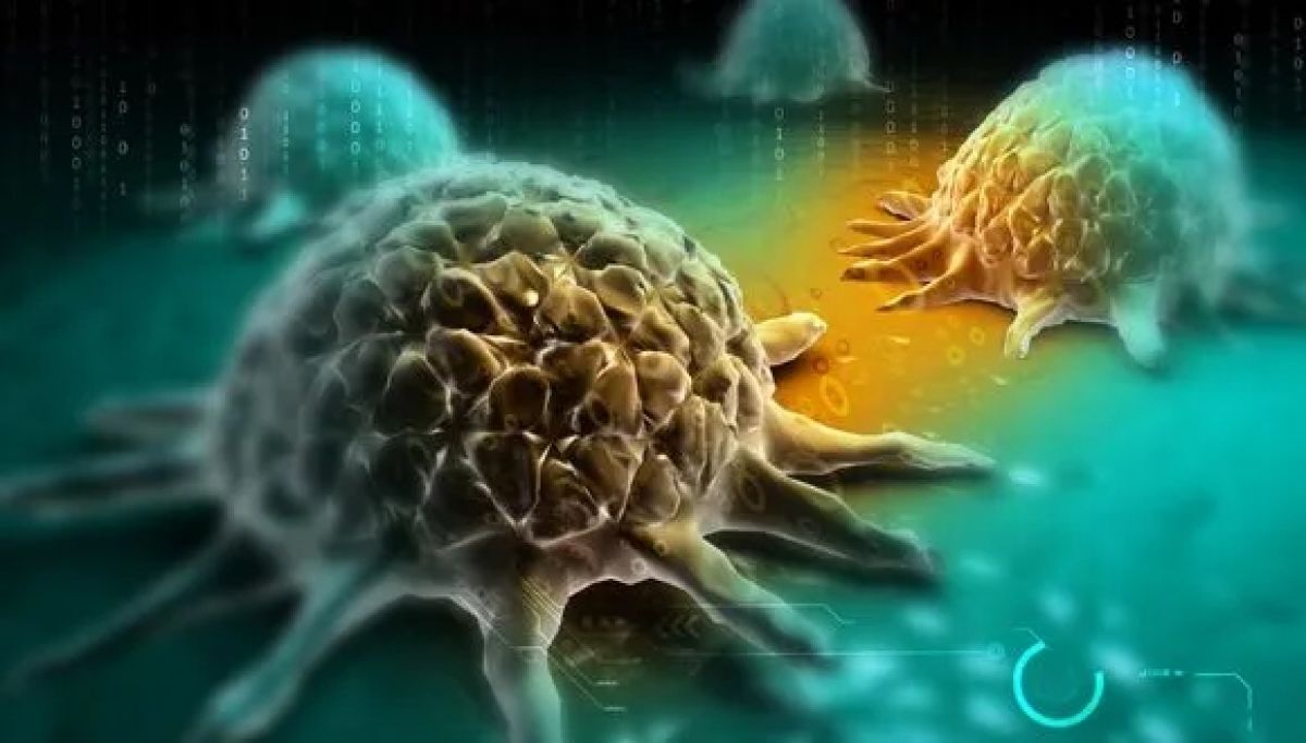
Heart Disease’s Cancer Link Unveiled

A Recent TAU Study Exposes the Connection Between Heart Disease and Cancer

Researchers at Tel Aviv University and the Leviev Cardiothoracic and Vascular Center at the Sheba Medical Center have found a mechanism that is responsible for increasing the risk of developing cancer among patients with heart disease: those small extracellular bubbles, or vesicles (sEVs), that are secreted from the sick heart to heal itself are released into the bloodstream – and promote the growth of cancer cells throughout the body. The researchers estimate that the important discovery may improve the protocols for treating heart disease so that clinicians also consider the increased risk of cancer. The study was funded by the Israel Cancer Association and the Israel Science Foundation.

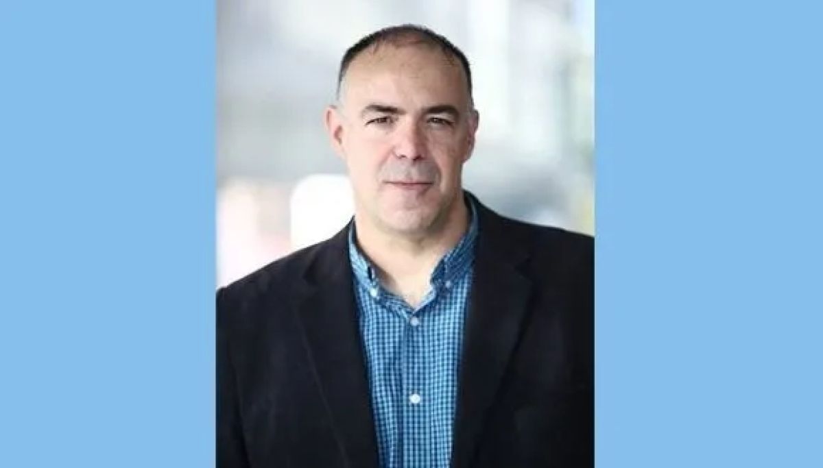
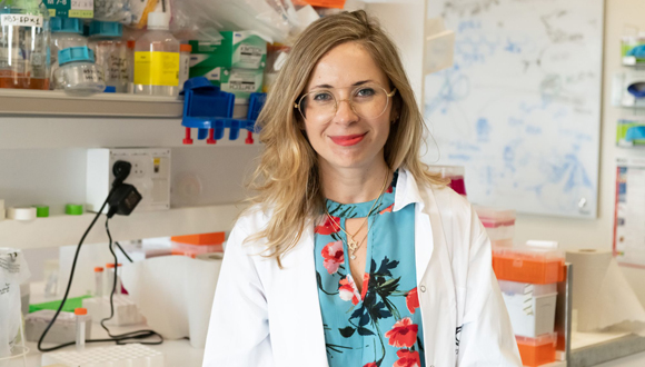
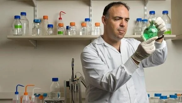
The research was conducted under the leadership of Prof. Jonathan Leor from the Neufeld Cardiac Research Institute, Faculty of Medical & Health Sciences at Tel Aviv University and the Taman Institute at Sheba’s Leviev Center and his student Tal Caller, a medical and research student at Tel Aviv University’s School of Medicine. The research was published in the important medical journal Circulation.

Heart Disease’s Silent Hand in Cancer’s Rise

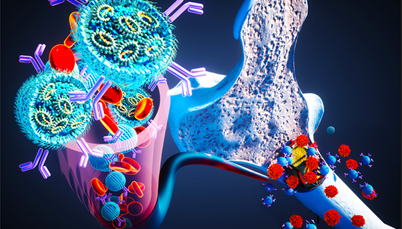
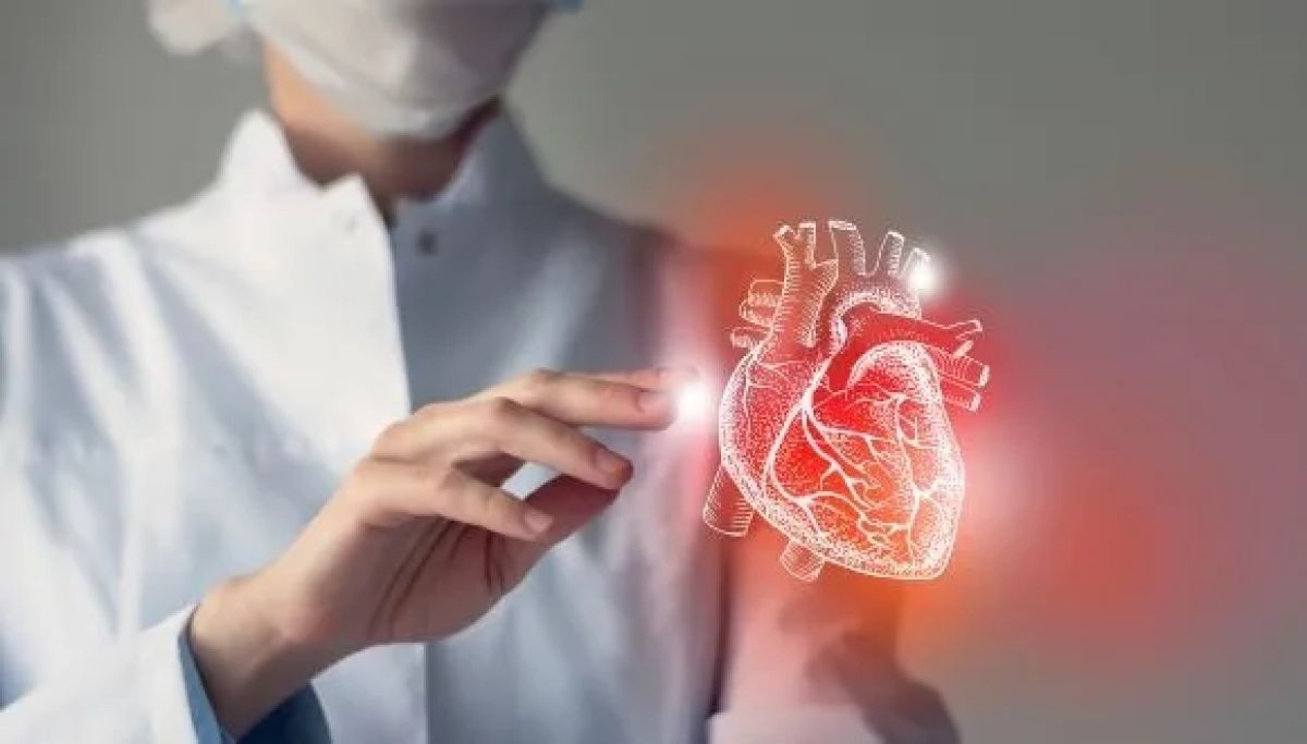
Caller explains: “In 2013, the Israeli cardiologist Tal Hasin showed for the first time that there is a connection between heart failure and cancer. Patients with heart disease are at a higher risk of developing cancer, and since heart disease is already a leading cause of death–first place in the US and second place in Israel – that means that many people are at risk. Our research revealed that the diseased heart secretes cancer-promoting factors, which we identified as small extracellular vesicles (sEVs). These are tiny particles wrapped in a simple membrane, which all cells secrete, However, due to heart damage, these vesicles are released in greater quantities and contain factors related to inflammation, healing, growth, creation of new blood vessels, and changes in the immune system. These vesicles move through the circulatory system and eventually reach the tumor or the pre-cancerous tissue”.

Caller adds, “Following an injury in the heart muscle and deterioration to heart failure, sEVs containing growth factors and small nucleic acid molecules that promote cell division are released. These sEVs contribute to the healing of the injured cardiac tissue. However, released from the injured heart, those vesicles move within the body’s circulatory system, eventually targeting cancerous growths”.

Prof. Jonathan Leor: “Many theories have been proposed to explain the increased risk of cancer in heart patients. They started with shared risk factors such as smoking, diabetes, and obesity and ended with a single protein or molecule. We showed for the first time that the diseased heart secretes sEVs that contain thousands of different growth factors. These bubbles directly promote the growth of certain tumors and also modulate the immune system, making the body more vulnerable to tumor growth”.

To test their hypothesis, the researchers at TAU inhibited the formation of sEVs in animal models with heart disease and found that the risk of cancer decreases along with the inhibition of vesicle production. However, this is not a viable therapeutic option since inhibiting the production of the vesicles causes severe undesired side effects.

Disrupting Cancer’s Path

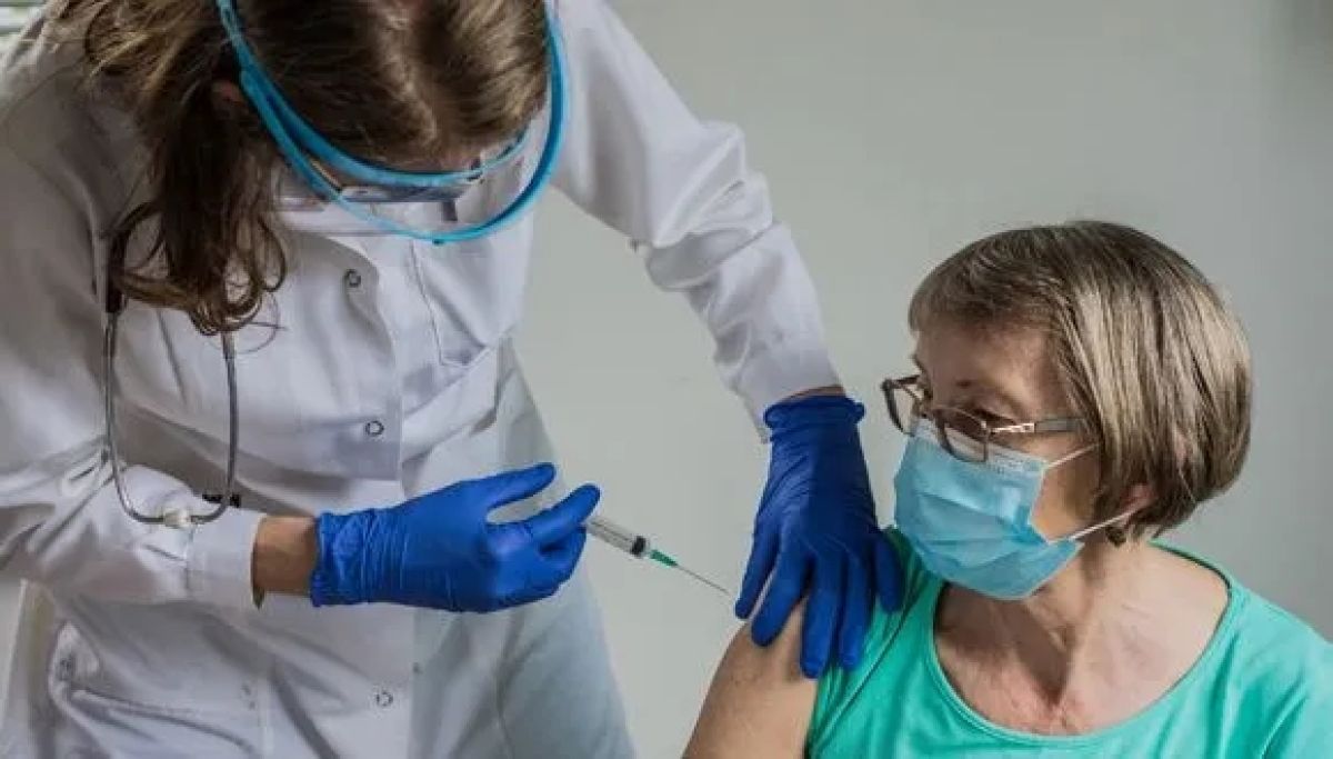
Prof. Leor: “When you systemically inhibit the formation of sEVs, you get less cancer – but you cause collateral damage along the way. That is why we tried a different strategy: treat the patient’s heart to reduce the damage to the cardiac tissue so that it secretes fewer sEVs. We used spironolactone, which is a well-known, old, and effective drug used to treat heart failure. We treated the animals with spironolactone at a very early stage of the disease and found that the heart secreted 30% fewer sEVs– and the cancerous tumors grew more slowly. Our experiment shows that it is possible to intervene in heart disease in a way that reduces the risk of cancer among heart patients”.

As for the clinical implications of the study, Caller is careful in his words: “It may be necessary to adjust the existing treatments for the heart so that they also consider the risk of cancer. In addition, it is possible to find biomarkers among heart patients that will indicate an increased risk of cancer since not all patients are at an increased risk. This is basic research, and much work is still required to unravel the connection between the two”.

Moshe Bar-Haim, CEO of the Israel Cancer Association, adds: “Thanks to public donations and designated funds, the research committee of the Israel Cancer Association examines and selects dozens of studies every year and funds researchers and doctors from research and treatment centers across Israel. From these studies, new methods were developed for the diagnosis, treatment, and rehabilitation of cancer patients. Research has no territorial boundaries, so every achievement in research here in Israel is an achievement for the entire world. We hope that the new research – that reveals that because of heart diseases, extracellular bubbles are secreted and increase the risk of cancer – will allow for immediate application in Israel and around the world for the benefit of accurate treatment for patients”.

Do Green Environments Help Heart Patients Live Longer?

Grass Is Greener, Lives Are Longer: Nature’s Impact on Heart Patients’ Health.

In a long-term study, unprecedented in its kind and scope, researchers from Tel Aviv University examined the association between a greener environment, which is most likely saturated with vegetation, and the mortality rate of coronary heart patients after undergoing bypass surgery – which is considered a traumatic event from both the physical and mental aspects. The study, which was carried out on thousands of patients who live all over the State of Israel and followed them over more than 10 years, found that the survival rate of bypass surgery heart patients who live in greener areas is significantly greater than those whose living environment is devoid of greenery. The study was conducted by Ph.D. student Maya Sadeh under the guidance of Prof. Rachel Dankner from the Department of Epidemiology and Preventive Medicine at the School of Public Health in the Faculty of Medical and Health Sciences and Prof. Alexandra Chudnovsky from the Porter School of Environment and Earth Sciences at TAU. Also participating: Nir Fulman, also from the Porter School, Nirit Agay and Arnona Ziv from the Gertner Institute for Epidemiology Research at Sheba Medical Center, Ilan Levy from the Ministry of Environmental Protection, and Prof. Michael Brauer from the University of British Columbia in Canada. The research was carried out with the support of the Environment and Health Fund and the Israel Science Foundation and was published in the prestigious journal Epidemiology.

Unique Study Involving 3,128 Heart Patients

Prof. Dankner: “The current study was based on a database we built at the Gertner Institute about 20 years ago for another study: 3,128 heart patients who underwent bypass surgery in seven medical centers in Israel, from Haifa to Beer Sheva, between the years 2004-2007. Using data from the Ministry of the Interior Affairs we found that 1,442 (46%) of them died of various causes by the year 2021. In this study, we wanted to examine to what extent (if at all) the life expectancy of heart patients after surgery is associated with the amount of green vegetation in their residential area”. For the study, the researchers cross-referenced the patients’ residential address data with data from NASA’s Landsat satellites, which photograph the Earth and can locate the color green with a very high resolution and within a range of up to 30×30 meters from the residential address – which allows identification of vegetation even within urban areas.

Nature’s Health Check

The researchers worked to accurately account for the amount of greenery in a radius of up to 300m around the address of each patient and placed this figure against the dates of death or survival of the patients over 14 years from the date of surgery. They performed a detailed statistical analysis of the data, including adjustments for a variety of variables, such as age, sex, ethnicity, socioeconomic status, urgency of the hospitalization (elective, semi-elective, or emergency surgery), living in the periphery/center, air pollution, and living distance from the Mediterranean Sea. About 90% of the research participants lived in urban areas, 80% in the coastal plain from the center to Haifa, 15% in the Jerusalem area, and 5% in the southern Beer Sheva area. Maya Sadeh: “We divided the residential addresses of the patients into three groups, according to the amount of vegetation in their surroundings, and found a clear significant association between a green environment and the survival of the patients – that is, how many years they continued to live after the operation. The results revealed that during the mean time of 12 years following the operation, the risk of mortality for those who lived in a very green environment was lower on average by 7% compared to those who lived in a non-green environment. We also found that the beneficial relationship is more pronounced among women, who made up 23% of the cohort, and were older at the time of the surgery (69.5 years old on average) compared to men (63.8 years )”.  
The researchers conclude: “In this study, we examined the survival of coronary heart patients after undergoing bypass surgery, and found that living in a greener environment is associated with better chances of survival. We hypothesize that there are a variety of reasons for this: in a green environment, people breathe cleaner air and engage in more physical activity, the atmosphere may be calmer, and the quality of life is better overall. The research findings may be particularly relevant to the current period in Israel: implying that exposure to a green environment may be a beneficial factor in recovering from trauma”.

TAU Receives $12.67M Grant for Medical Simulation Center

The Faculty of Medical and Health Sciences at Tel Aviv University Receives a $12.67 Million Grant from the Helmsley Charitable Trust to Create a Medical Simulation Training Center.

TEL AVIV, Israel & NEW YORK—A $12.67 million lead grant from The Leona M. and Harry B. Helmsley Charitable Trust will support the creation of the Helmsley Medical Simulation Center (“Center”) at the Faculty of Medical and Health Sciences at Tel Aviv University. The Center will provide student training for a new generation of skilled, experienced, and compassionate medical, dental, and healthcare professionals. The skills learned through simulation trainings will lead to increased patient safety and improved health outcomes. The grant will be used to plan, build, equip, and begin operating the Center in an existing building on Tel Aviv University’s campus. The term “medical education” encompasses many goals, but its key challenge is to teach medical professionals the clinical expertise and communication skills they need to successfully treat patients while managing complex interactions with patients, families, and team members. Managing these sensitive interactions appropriately is critical for ensuring high-quality care and patient safety. In addition, students must practice providing care without causing harm or putting patients at risk. Medical simulation training centers have thus become the standard of care worldwide and a synonym for excellence. The Center will be a dynamic and vibrant facility in use from morning to night by 1,400 medical and dental students, 3,000 MDs receiving continuing education at the Center for Continuing Medical Education at Tel Aviv University, and an additional 500 nursing students, physical therapists, occupational therapists, and more. With Israel facing an acute shortage of medical staff, the Center is a timely initiative, enabling the Faculty of Medical and Health Sciences to expand medical training opportunities by revamping medical curricula, prioritizing critical thinking and problem-solving skills, and promoting a holistic patient care approach. The Center will provide comprehensive, high-quality, safe, and culturally sensitive educational services by employing state-of-the-art technologies in a teaching and research environment that is innovative and collaborative. Under the supervision of medical faculty members, the Center will include mock doctor-patient encounters featuring either life-like mannequins or actors as patients, and assessment that includes video-based feedback. Such activities will allow trainees to hone their communication and technical skills in a safe and controlled learning environment that is also dynamic and interactive. Such an environment can help improve student performance, engagement, and retention, ultimately leading to better outcomes for students and later for patients. The Center will implement extended and virtual reality technology, as well as extensive multimedia platforms, with a strong research component.  
“We support the people of Israel, and we are committed to supporting Israel’s world-class healthcare services,” said Sandor Frankel, a Trustee of The Helmsley Charitable Trust.
  “The Faculty of Medical and Health Sciences at Tel Aviv University is a powerhouse of teaching and research in medicine and the health sciences, and we recognize the impact the University will have through providing state-of-the-art medical training for a new generation of students, physicians, nurses, occupational therapists, and other health professionals.” Helmsley aims to expand healthcare services so that everyone in Israel has access to quality healthcare no matter where they live, strengthen Israel’s contributions to basic scientific, technological, and medical research, and promote global understanding and appreciation of Israel and its people. Previous grants from Helmsley have supported Tel Aviv University’s research on nanomedicines for personalized “theranostics” for cancer, cardiovascular, and inflammatory diseases, and research on advanced communications technology. Tel Aviv University President, Prof. Ariel Porat, said, “We are deeply grateful for this vote of confidence by the Helmsley Charitable Trust in our medical faculty, which is the largest and leading one in Israel and among the most advanced in the world. With the visionary Helmsley gift, we will be able to build a major facility that is comprehensive, collaborative and innovative, and that will bring our medical training to new heights for the benefit of the Israeli people.”  
Prof. Karen Avraham, Dean of the Faculty of Medical and Health Sciences, commented: “Our remarkable team has worked tirelessly in the recent months to bring this dream to reality, Inspired by our former Dean, Prof. Ehud Grossman. At a time when medical and health care is on the verge of transformations in AI and gene and cell therapy, providing competency and compassion remains a priority.”
 

About the Helmsley Charitable Trust

The Leona M. and Harry B. Helmsley Charitable Trust aspires to improve lives by supporting exceptional efforts in the U.S. and around the world in health and select place-based initiatives. Since beginning active grantmaking in 2008, Helmsley has committed $4.5 billion for a wide range of charitable purposes, including over $605 million for projects in Israel. For more information on Helmsley and its programs, visit helmsleytrust.org.

About Tel Aviv University

Tel Aviv University (TAU) – Israel’s largest and most comprehensive institution of higher learning – is home to over 30,000 students studying in nine faculties and over 125 schools and departments across the spectrum of sciences, humanities, and the arts. Situated in Israel’s cultural, financial, and technological capital, TAU shares Tel Aviv’s unshakable spirit of openness and innovation – and boasts a campus life as dynamic and pluralistic as the metropolis itself. Tel Aviv the city and Tel Aviv the university are one and the same – a thriving Mediterranean center of diversity and discovery.

Breaking the MedTech Glass Ceiling

An alumna of TAU’s program for young women entrepreneurs is developing a device that could revolutionize heart disease treatment.

Israel’s reputation as “Startup Nation” is well-deserved, but its tech industry suffers from a lack of gender diversity. Hoping to bring in a wealth of untapped potential, Tel Aviv University’s Entrepreneurship Center created a semester-long hands-on workshop for women to help bring their startup ideas to fruition with the help of experienced mentors. One such project is Symbiosis CM, founded by student-and-mentor team Dr. Shira Burg and Varda Badet, which is creating a medical device that personalizes treatment of heart disease. The startup has raised $1.9M as of last year and won a number of competitions and grants including the Deep Tech Track of the Coller Startup Competition and a grant from the Israeli Innovation Authority. The Problem  Originally trained as a veterinarian, Shira Burg worked for seven years alongside a small animal cardiologist in Israel, doing catheter procedures on dogs with congenital heart valve disease. There, she learned that one of the most common heart diseases, mitral valve disease, is difficult to treat because valve replacements are not fitted to individual heart sizes and replacement surgeries are invasive and costly. The disease occurs in aged dogs—and aged humans—in almost exactly the same way. During this period, she began working with a surgeon who was attempting to create a mitral valve prosthetic, but discovered just how much heart anatomies differ from person to person. “You can’t take something rigid and standard and expect it to fit every heart,” she says. “I realized that though we have personalized medicine in many fields such as drugs, diet, and genetics, we don’t have much personalization for medical devices. I saw an opening there for an improved mitral valve solution.”  
Shira Burg: “I realized that though we have personalized medicine in many fields such as drugs, diet, and genetics, we don’t have much personalization for medical devices. I saw an opening there.”
  Burg decided to pursue a PhD in cardiac electrophysiology at TAU’s Faculty of Medical and Health Sciences at the lab of Prof. Bernard Attali. There, she continued to think about her idea for a better mitral valve device. In 2020, she heard about a new workshop, Yazamiyot, for female graduate students at TAU’s Entrepreneurship Center. “I realized that if I could get accepted, I might be able to do something with my idea.” After a rigorous application process involving three separate interviews, Burg was one of 30 women admitted to the first course. The Course  Yazamiyot, meaning “female entrepreneurs”, is an accelerator program targeting women master’s and PhD students. Participants work in small groups over one semester to establish a startup initiative in a supportive and empowering environment and are mentored through the process by successful women with industry experience. Burg’s mentor, TAU alumna Varda Badet, would go on to become her business partner. “With Varda, something just stuck. We had a connection,” Burg says. Badet came from the finance sector, having served as the EVP at Bank Leumi for Risk Management during her many years in the field. Hoping to share her knowledge and connections, she took some courses to become a mentor at the Entrepreneurship Center. “There I found Shira, a very special entrepreneur. Every unsolved problem, she investigates and explores until it is solved. She is very motivated and ambitious, and I could see she was someone I wanted to work with.”  
Yair Sakov: “At Tel Aviv University, we feel it is our responsibility to help drive change within society. Our mission at the Center is to empower students to come up with solutions to global challenges.”
  During the semester, participants were brought face to face with the market realities that would determine whether their ideas were commercially feasible. They were taught how to inform themselves on target demographics and competitors, present their ideas to experts and the public, build a team, and more. They also heard from industry experts, from whom they could ask advice on their individual projects, and successful female entrepreneurs. Yair Sakov, founder and managing director of the TAU Entrepreneurship Center, says: “At Tel Aviv University, we feel it is our responsibility to help drive change within our society. Our mission at the Center is to empower students to come up with solutions to global challenges by helping them develop an entrepreneurial mindset and by giving them tools and resources. In particular, we aim to uplift underrepresented communities, including women, who are still marginalized in the startup ecosystem. The Yazamiyot program was created to address that gender gap, and nearly every cohort since its inception has contributed to the establishment of new woman-led startups.” Burg credits the workshop with giving her the tools to make her idea for a personalized heart valve device a reality, but says the most important thing she gained was her connection with Badet. Aside from teaching necessary business finance skills, Badet brought in resources in the form of her many industry connections and financial backing. “As a mentor, you must believe in your entrepreneur completely and be ready to stick out the tough process with them. This product is worth that perseverance because it could save so many lives,” says Badet. The Company  There is indeed great life-saving potential in addressing the lack of effective mitral valve disease solutions: in the US alone about 70% of the 4 million patients with the condition are unable to get proper treatment. Additionally, hospitals pay about $40 billion a year in treatment costs, with over 90% rehospitalization rates due to heart failure. Many companies have tried to address the problem, but their products are not widely approved and do not fit more than 20% of heart anatomies. In a novel move, Symbiosis CM is developing a docking system that allows for real-time valve adjustment per an individual’s heart structure and minimally invasive procedures to treat the disease. The system is compatible with valves already on the market, making it accessible and opening the door to collaboration with other companies. The startup team already has a working prototype in preclinical trials on lab models.   A sample image of the heart valve docking system created by Symbiosis CM Burg happily notes that her original plan to develop a solution for dogs, ideated during her time as a veterinarian, may one day also become reality: the product is small enough to work on animals as well as people. The management side of things is also on track for success: Burg and Badet recently brought on a new CEO, Amir Weisberg, who has behind him 35 years of entrepreneurial experience, three exits in the medical field and an IPO on NASDAQ. “He was actually retired when we approached him, but after we presented to him, he decided to come out of retirement and sign on full-time to the company,” says Burg. The Need for Women in Industry  
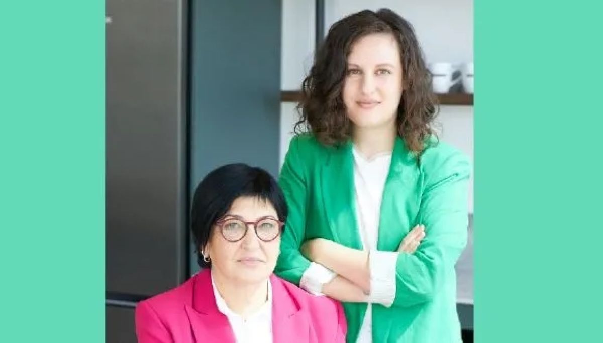
Both Burg and Badet note that the Yazamiyot course is essential because of the difficulties of succeeding in the startup industry as a woman. Says Burg, “It’s a male-dominated world. We need to say it out loud. Especially in the spaces where I work, in the medical field and cardiology, it’s mostly men. When I came to specialists and investors, they would see a young woman and decide before I started speaking that I wasn’t to be taken seriously. That is why it is so important to push more women into entrepreneurship in general: so that people no longer question what we’re doing there.” Adds Badet, “it makes me so glad to see and to help more women break into entrepreneurship.”  
Shira Burg: “In Israel, female entrepreneurship is an unpolished diamond.”
  “Israel has many undiscovered talents,” says Burg. “Female entrepreneurship, especially, is an unpolished diamond. If we expand programs such as Yazamiyot, amazing things will come from it.” About the Entrepreneurship Center  Four the last four years, Tel Aviv University’s Entrepreneurship Center has provided students from across campus with the knowledge, tools, strategies and opportunities to create business and social ventures. It connects them with alumni, industry, government agencies and NGOs to generate and develop the next world-changing ideas. Over the Center’s first years of operations, it has achieved the following:   · 100 entrepreneurship courses, events and programs · 12,000 student participants · 92 startups ·$155 million in VC capital ·190 top industry mentors, most of them TAU alumni By gradually expanding activities, the Center expects its to reach 4,500 students per year by 2029.    

Summer Glow: How Sun Exposure Boosts Fertility in Women Ages 30-40

Exposure to The Sun’s UV Radiation May Have a Positive Effect on Fertility in Women Aged 30-40.

A research team from Tel Aviv University and the Sheba Medical Center at Tel Hashomer investigated seasonal fluctuations in AMH (anti-Müllerian hormone) levels. Their pioneering study revealed that during the summer, women of a late reproductive age — between the ages of 30 and 40 — experience increased secretion of the hormone from their ovaries.   This phenomenon is suggested to be attributed to heightened exposure to ultraviolet (UV) radiation from the sun. The groundbreaking research was led by Prof. Carmit Levy of the Department of Human Genetics and Biochemistry, in a team effort of Ph.D. student Roma Parikh and Prof. Yftach Gepner of the School of Public Health, all from the Faculty of Medicine at Tel Aviv University and Dr. Ruth Percik from the Institute of Endocrinology at Sheba Medical Center. The results of the study were published in the journal Steroids.   “The ovaries secrete the anti-Müllerian hormone, and its level in the bloodstream is linked to ovarian function,” explains Dr. Percik. “While the hormone level is specific to an individual woman at a given point in time, and does not provide a definitive assessment of the status of her fertility, evaluating its value, trend, and comparison to the age group is the best indicator of fertility that we have. For this reason, every woman who wants to get pregnant, or is trying to, is sent for an AMH test. In Israel, all of these tests are directed to the central laboratory in Sheba. Our research group investigated the seasonal variability of the AMH tests to gauge how the ovaries respond to UV radiation”.  

Moderation in Sun Exposure: Key to Fertility Health

The researchers compared the AMH results of 2,235 Israeli women to the recorded levels of UV radiation. For younger women, aged 20-29, no statistical relationship was found between UV exposure and AMH level. On the other hand, among older fertile women, aged 30 to 40, a statistically significant seasonal pattern emerged: These women, whose egg reserves are in decline, responded positively to sun exposure.  
“Based on our prior studies, we can affirm that sun exposure increases metabolism, as well as sexual appetite and behavior, and (at least in animal models) enlarges the ovaries and extends the estrus period” explains Prof. Levy.
  “This is a preliminary, pioneering human epidemiological study, and we need to be cautious about inferring a causal relationship between fertility in women and exposure to UV radiation. Humans are not the same as mice. However, we are also animals, our hairless nature makes us even more sensitive to solar radiation. Our research suggests that the female reproductive system is indeed more fertile in the summer, but we still have no information on the mechanism or actual success rates”, she continues. Particularly interesting is the absence of this effect among younger women in their 20s. According to Dr. Percik, this may be attributed to the ample egg reserve found in young women. “Based on my interpretation of the findings, women at the onset of their reproductive age are less in need of signals from the sun, which affect hormonal pathways that have not yet been sufficiently studied. They are less impacted or dependent on the forces of nature in the context of fertility. In contrast, older ovaries need optimal environmental factors to function. In fact, this effect was even more pronounced among women aged 35 and older. Of course, there are caveats: Exposure to the sun’s UV radiation should always be done in moderation, and further research is required to determine whether such exposure actually helps fertility, and how much exposure is needed”.

Are We Close to Ending Alzheimer’s Memory Loss?

TAU Researchers Successfully Prevent Memory Deterioration in Alzheimer’s in an Animal Model.

In 2022, a team of researchers from the laboratory of Prof. Inna Slutsky from the and the at TAU uncovered a pathological brain phenomenon in an animal model that precedes the first appearance of Alzheimer’s disease symptoms by many years. This is an increased activity in the hippocampus during the states of anesthesia and sleep, which results from damage to the mechanism that stabilizes the neural network.

Breakthrough in Alzheimer’s Study

In the current study published in Nature Communications, Prof. Slutsky’s laboratory team, in collaboration with the Safra Center for Neuroscience at the Hebrew University, found that suppression of neuronal activity in a small nucleus in a specific area of the thalamus (which regulates sleep states) caused a decrease in pathological activity in the hippocampus and prevented the deterioration of the memory in Alzheimer’s in an animal model. The researchers hope that their research will speed up the start of clinical trials in humans, lead to progress in the fields of early detection and prevention of the onset of dementia symptoms in Alzheimer’s disease, and in the field of treating cognitive impairments caused by surgery (POCD – Postoperative Cognitive Dysfunction).

Doctoral Student Shiri Shoob

Doctoral Student Shiri Shoob

“As early as 10-20 years before the appearance of the familiar symptoms of memory impairment and cognitive decline, physiological changes slowly and gradually occur within the patients’ brains,” explains the doctoral student who led the study, Shiri Shoob. “There is an accumulation of amyloid-beta deposits and abnormal accumulations of tau protein, a decrease in the volume of the hippocampus, and more. Moreover, about 30% of the people who were found to have a pathology typical of Alzheimer’s disease at postmortem did not develop the typical symptoms of the disease during their lifetime. It seems, then, that the brain has an, admittedly limited, ability to protect itself from the damage of the disease.”

Uncovering Alzheimer’s Protective Mechanisms

The research focused on finding those protective mechanisms that the brain has against the damage from the disease. The researchers found that during sleep – and especially during sleep as a result of general anesthesia – the early symptoms of Alzheimer’s disease, which appear many years before the symptoms of dementia, could be more easily identified. Prof. Inna Slutsky: “Anesthesia reveals pathophysiology in the brain activity in the animal model. We think that there are mechanisms that compensate for that same pathology while awake and thus prolong the pre-symptomatic period of the disease”.

Prof. Inna Slutsky

Prof. Inna Slutsky

The researchers identified hyperactivity in the hippocampus – “silent seizures,” which look like an epileptic seizure in terms of brain activity, but do not appear externally – in in an animal model of Alzheimer’s disease.This, compared to the reduced activity in the healthy hippocampus during sleep and anesthesia. To examine potential treatment and prevention measures, the researchers tried a variety of methods, but mainly focused on deep brain stimulation (DBS) using electrical signals to the nucleus reuniens – a small nucleus in the brain that connects the affected hippocampus and the thalamus, which is responsible for sleep regulation.

“When we tried to stimulate the nucleus reuniens at high frequencies, as is done in the treatment of Parkinson’s, for example, we found that it worsened the damage to the hippocampus and the silent epileptic seizures,” said Shoob, “only after changing the stimulation pattern to a lower frequency were we able to suppress the seizures and prevent cognitive impairment. We showed that the nucleus reuniens could completely control these seizures. We could increase or decrease the seizures by stimulating it”.

Decoding Alzheimer’s

Prof. Slutsky adds: “Epidemiological studies indicate a link between aging and a phenomenon called POCD – cognitive problems that arise after surgery under general anesthesia. In young people, the symptoms usually pass very quickly, but in older people, the chance of cognitive impairment increases and it may last a long time. Our research indicates a potential mechanism underlying the phenomenon. We found that suppressing the thalamic nucleus reuniens – by pharmacological or electrical means – successfully prevented both pathological activity in the hippocampus during anesthesia and cognitive impairment following anesthesia.

In addition, we identified a relationship between certain pathological activity in the hippocampus during anesthesia in the presymptomatic phase of Alzheimer’s to memory problems in a more advanced stage of the disease. This indicates a potential for predicting the disease in the dormant state, before the onset of cognitive decline”.

The leader of the study, Shiri Shoob, added: “We saw that no matter what means we used, when we inhibited the neural activity in the nucleus, we also measured a decrease in the pathological activity in the hippocampus during anesthesia”.

The research was led by PhD student Shiri Shoob, with the participation of Nadav Buchbinder, PhD student Ortal Shinikamin, Halit Baeloha, Dr. Tomer Langberg, Dr. Daniel Zarhin, Dr. Ilana Shapira and Dr. Gabriella Braun from Prof. Inna Slutsky’s lab and in collaboration with Dr. Naomi Habib and Or Gold from the Hebrew University.

Destroying Cancer: new drug delivery system containing RNA therapy can target cancer cells in bone marrow

Researchers from Tel Aviv University develop nanoparticles containg RNA molecules,  similar to those used in the COVID-19 vaccine, that inhibit the ability of cancer cells to divide.

Researchers at Tel Aviv University destroyed 90% of the multiple myeloma blood cancer cells under laboratory conditions, and 60% in human tissues taken from patients at Rabin Medical Center (Belinson Hospital), using an RNA-based drug delivered to the cells by targeted lipid nanoparticles.

The researchers developed lipid-based nanoparticles (similar to those used in the COVID-19 vaccine) containing RNA molecules that silence the gene CKAP5, encoding cytoskeleton-associated protein 5. With this protein inhibited the cancer cell is unable to divide, which essentially kills it. To avoid damaging noncancerous cells, the nanoparticles were coated with antibodies that guided them specifically to the cancer cells inside the bone marrow.

The breakthrough was achieved by a group of researchers from Tel Aviv University and the Rabin Medical Center, led by Prof. Dan Peer, a pioneer in the development of RNA therapeutics and Head of the Nanomedicine Laboratory at the Shmunis School of Biomedicine and Cancer Research, also serving as TAU’s VP R&D, and by PhD student Dana Tarab-Ravski. The results were published in the leading journal Advanced Science.


Cover image for Advanced Science (credit: Ella Maru Studio)


“The targeted drug delivery system we developed is the first to effectively reach cancer cells inside the bone marrow.”


Dana Tarab-Ravski explains: “Multiple myeloma is a blood cancer usually found in the older population. While most blood cancers appear in the blood stream or lymph nodes and spread from there to the rest of the body, multiple myeloma cells appear and form tumors inside the bone marrow – and are therefore very hard to reach.”

The study’s findings are very encouraging: under laboratory conditions, where cells are grown in flasks, the nanoparticles developed by the researchers eradicated about 90% of the cancer cells. At the second stage, the new treatment was tested on cancer samples taken from multiple myeloma patients at the haemato-oncological ward of the Rabin Medical Center. The success rate in these samples was 60%. Testing the ability of the nanoparticles to reach the bone marrow in an animal model, the researchers found that after a single injection the RNA had penetrated to 60% of the multiple myeloma cancer cells in the bone marrow. Lastly, examining the therapeutic effectiveness of the nanoparticles in the animal model resulted in eradication of two thirds of the cancer cells, and the animals showed significant improvement in all clinical indicators.

“People with multiple myeloma suffer from severe pain in their bones, as well as anemia, kidney failure, and a weakened immune system,” says Tarab-Ravski. “There are many possible treatments for this disease, but after a certain period of improvement most patients develop resistance to the therapy and the disease relapses even more aggressively. Therefore, there is a constant need for developing new treatments for multiple myeloma. RNA-based therapy has a great advantage in this case because it can be developed very quickly. By simply changing the RNA molecule a different gene can be silenced each time, thereby tailoring the treatment to the progression of the disease and to the individual patient. The challenge in these treatments is to reach the right cells. Today RNA therapeutics are approved for treating a genetic liver disease and for vaccines injected into the muscle, as we saw with the COVID-19 vaccines. The drug delivery system we developed is the first that specifically targets cancer cells inside the bone marrow, and the first to show that silencing the expression of CKAP5 gene can be used to kill blood cancer cells.”

Prof. Dan Peer: “Our technology opens a new world for selective delivery of RNA medications and vaccines for cancer tumors and diseases originating in the bone marrow.”

The clinical team included Dr. Tamar Berger, Dr. Iuliana Vaxman and Prof. Pia Raanani from the Institute of Hematology, Rabin Medical Center, Beilinson Hospital. The research was funded by the the Boaz and Varda Dotan Hematologyoncology Center at Tel-Aviv University and by Lewis Trust for Blood Cancer Research awarded to D.P.

Stress Makes Vaccines Less Effective

Researchers at Tel Aviv University find correlation between behavioral stress and vaccine effectiveness.

Researchers at Tel Aviv University demonstrated for the first time that there is a significant link between behavioral stress and the effectiveness of vaccines. They found that acute stress in lab models 9-12 days after vaccination increases antibody response to the vaccine by 70% compared to the unstressed control group. This, however, comes at the price of reduced antibody breadth, which results in diminished protection against the pathogen’s variants.


“Our study was the first to investigate the possible effects of acute stress. We found that this mental state has a dramatic impact – not only on the vaccine’s effectiveness, but also on how it works.” – Dr. Natalia Freund


The “Dramatic Impact” of Stress

The study was carried out in Tel Aviv University and led by Ph.D. student Noam Ben-Shalom from the lab of Dr. Natalia Freund at the Faculty of Medicine and Ph.D. student Elad Sandbank from the Neuro-immunology Lab of Prof. Shamgar Ben-Eliyahu at The School of Psychological Sciences and the Sagol School of Neuroscience. The paper was published on July 6th in the leading scientific journal Brain, Behavior, and Immunity.

Dr. Freund explains: “In this study we examined, for the first time, the correlation between stress and the body’s ability to develop an immune response following vaccination. The prevailing assumption is that the effectiveness of a vaccine is determined mainly by its own quality. However, over the years, professional literature has reported influences of other factors as well, such as the age, genetics, and microbiome of the outcomes of vaccination. Our study was the first to investigate the possible effects of acute stress. We found that this mental state has a dramatic impact – not only on the vaccine’s effectiveness, but also on how it works.” 

Classical ‘Fight or Flight’ Response

Acute stress is a mental state caused by immediate threat (either real or imagined), involving the secretion of adrenaline and stimulation. In this study, Dr. Freund and her colleagues vaccinated mice with two different vaccines: the model protein Ovalbumin and a fragment of the SARS-CoV-2 spike protein also used in the COVID-19 vaccine. Nine days later, just as the adaptive immunity became active and the production of antibodies began, the mice were subjected to a widely used behavioral paradigm simulating acute stress. Two and a half weeks after exposure to stress, namely 30 days after vaccination, the level of antibodies in the blood of vaccinated animals that had experienced stress was 70% higher compared to the control group. This phenomenon was observed in animals vaccinated with either type of vaccine.

At the same time, the researchers discovered that the immune system of the animals that had experienced stress was not cross reactive to variants of the protein used in the vaccine. In other words, following stress the immune system was focused entirely on the original vaccine, showing no response to proteins that were only slightly different – such as variants of concern (VOC) of SARS-CoV-2.


Dr. Natalia Freund


“In general, the purpose of vaccination is not only protection against a specific pathogen, but also creating a long-lasting immunological memory for protection against future mutations of that pathogen.” – Dr. Natalia Freund


“Initially, we were surprised to find out that the response to the vaccine was much more effective in animals that had experienced stress,” says Dr. Freund, “we would have assumed just the opposite – that stressful situations would have a negative impact on the immune system. Nevertheless, with both types of vaccines, we observed a stronger immune response after stress, both in the blood and in B cells (the lymphocytes that produce antibodies) derived from the spleen and lymph nodes of the immunized mice. The enhancement of the antibodies’ activity following stress was mediated by the cellular receptor that identifies adrenaline – the beta2 adrenergic receptor. When we blocked this receptor, either pharmacologically or by means of genetic engineering, the effects of stress were completely eliminated. On the other hand, to our great surprise, the breadth of the immune response generated by the vaccine was reduced by about 50% following stress. In general, the purpose of vaccination is not only protection against a specific pathogen, but also creating a long-lasting immunological memory for protection against future mutations of that pathogen. In this sense, the vaccines appeared to lose much of their effectiveness after exposure to stress.”

According to the researchers, this is in fact a classical ‘fight or flight’ response, however this time demonstrated at the molecular level. During stress, the immune system produces large quantities of antibodies and stronger antibodies, to address the immediate infection, and this large energetic investment in the here and now comes at the expense of future immunological memory.

Does It Apply to Humans?

Dr. Freund adds: “In the second part of the study we wanted to test whether humans also display the post-stress immune impairment observed in vaccinated mice. For this purpose, we cultured B cells obtained from blood of people who had contracted COVID-19 in the first wave. We then induced stress in these cultures using an adrenaline-like substance that stimulates the beta2 adrenergic receptor, that was identified by us in the first part of the study as a mediator of the response to stress in cells that produce antibodies in mice. B cells express a very high level of these receptors, but until now the receptors’ role in producing antibodies was not known. In fact, it was unclear why these cells need the ability to respond to adrenaline.”

“We discovered that just like in mice, human cells also exhibit a zero-sum game between the intensity and breadth of the immune response. When the adrenaline receptor is activated during stress, the entire immune system is stimulated, generating antibodies that are 100-fold stronger than antibodies produced in cells that had not undergone stress. But here too, the response was narrower: the diversity of antibodies was reduced by 20-100%, depending on the individual from whom the cells were taken.


“Stress 9 to 12 days after vaccination, at the time when B cells are generating high affinity antibodies, enhances short-term immunity and damages long-term memory.” – Dr. Natalia Freund


RNA sequencing of the cells in which the beta 2 adrenergic receptor was activated, compared to regular cells, indicated that the receptor’s activation caused antibody-producing cells to work at maximum capacity (by activating the PI3 kinase protein and phosphorylation of AKT) – at the expense of antibody breadth and diversity.”

“From the evolutionary perspective,” concludes Dr. Freund, “stress can be caused by different factors. We tend to think of mental stress, but physical illness also causes a form of stress. When the body contracts a virus or bacteria it experiences stress, and signals to the immune system that the top priority is getting rid of the pathogen, while investing energy in long-term immunological memory is a second priority. Therefore, stress 9 to 12 days after vaccination, at the time when B cells are generating high affinity antibodies, enhances short-term immunity and damages long-term memory.”

Researchers Induce Cancer Cell “Suicide”

Tel Aviv University’s breakthrough study unleashes self-produced toxin, targeting and eliminating cancer cells with impressive results.

For the first time in the world: researchers at Tel Aviv University encoded a toxin produced by bacteria into mRNA (messenger RNA) molecules and delivered these particles directly to cancer cells, causing the cells to produce the toxin – which eventually killed them with a success rate of 50%.


“Our idea was to deliver safe mRNA molecules encoded for a bacterial toxin directly to the cancer cells – inducing these cells to actually produce the toxic protein that would later kill them. It’s like placing a Trojan horse inside the cancer cell.” – Prof. Dan Peer


“It’s like placing a Trojan horse inside the cancer cell”

The groundbreaking study was led by PhD student Yasmin Granot-Matok and Prof. Dan Peer, a pioneer in the development of RNA therapeutics and Head of the Nanomedicine Laboratory at The Shmunis School of Biomedicine and Cancer Research, also serving as TAU’s VP R&D. The study’s results were published in Theranostics.

Prof. Peer explains: “Many bacteria secrete toxins. The most famous of these is probably the botulinum toxin injected in Botox treatments. Another classic treatment technique is chemotherapy, involving the delivery of small molecules through the bloodstream to effectively kill cancer cells. However, chemotherapy has a major downside: it is not selective, and also kills healthy cells. Our idea was to deliver safe mRNA molecules encoded for a bacterial toxin directly to the cancer cells – inducing these cells to actually produce the toxic protein that would later kill them. It’s like placing a Trojan horse inside the cancer cell.”

Prof. Dan Peer

Impressive Results

First, the research team encoded the genetic info of the toxic protein produced by bacteria of the pseudomonas family into mRNA molecules (resembling the procedure in which genetic info of COVID-19’s ‘spike’ protein was encoded into mRNA molecules to create the vaccine). The mRNA molecules were then packaged in lipid nanoparticles developed in Prof. Peer’s laboratory and coated with antibodies – to make sure that the instructions for producing the toxin would reach their target, the cancer cells. The particles were injected into the tumors of animal models with melanoma skin cancer. After a single injection, 44-60% of the cancer cells vanished.  


“With a simple injection to the tumor bed, we can cause cancer cells to ‘commit suicide’, without damaging healthy cells. Moreover, cancer cells cannot develop resistance to our technology as often happens with chemotherapy – because we can always use a different natural toxin.” – Prof. Dan Peer


“In our study, the cancer cell produced the toxic protein that eventually killed it,” says Prof. Peer. “We used pseudomonas bacteria and the melanoma cancer, but this was only a matter of convenience. Many anaerobic bacteria, especially those that live in the ground, secrete toxins, and most of these toxins can probably be used with our method. This is our ‘recipe’, and we know how to deliver it directly to the target cells with our nanoparticles. When the cancer cell reads the ‘recipe’ at the other end it starts to produce the toxin as if it were the bacteria itself and this self-produced toxin eventually kills it. Thus, with a simple injection to the tumor bed, we can cause cancer cells to ‘commit suicide’, without damaging healthy cells. Moreover, cancer cells cannot develop resistance to our technology as often happens with chemotherapy – because we can always use a different natural toxin.”

Other contributors to the study included: Dr. Assaf Ezra, Dr. Srinivas Ramishetti, Dr. Preeti Sharma Dr. Gonna Somu Naidu and Prof. Itai Benhar, Head of the Antibody Engineering Lab at the Shmunis School of Biomedicine and Cancer Research at TAU. The study was funded by the Shmunis Family Foundation for Biomedicine and Cancer Research.

Metabolomics – A New Frontier in Preventive Medicine

Tel Aviv University’s new Metabolite Medicine Division at the BLAVATNIK Center for Drug Discovery poised to revolutionize the field.

Even the simplest blood tests of today – which monitor about 20 substances in our body – have powerful predictive and diagnostic power. For example, high cholesterol suggests possible heart trouble, and abnormal glucose could indicate pre-diabetes.

Now imagine that routine and low-cost bloodwork could check for thousands of compounds all at once, as well as calculate the balance between them. Such a real-time status check would provide doctors with unparalleled knowledge for diagnosing patients and creating personalized profiles for the most effective treatment of disease.


“Metabolomics is poised to revolutionize the field of preventive medicine. It holds tremendous potential (…) not only for detecting diseases but also for enabling individuals to proactively monitor their overall physiological well-being even before the onset of illness.” – Prof. Ehud Gazit


The Super Blood Test

We are entering such an era at TAU’s new Metabolite Medicine Division at the BLAVATNIK CENTER for Drug Discovery. This suite of labs is the most advanced at an Israeli university for the emerging science of metabolomics – the study of small molecules called metabolites that our bodies produce every second of our lives as part of ongoing cell processes. Sometimes, all that is required is to identify the one metabolite culprit that is throwing the body off balance.

“Metabolomics is poised to revolutionize the field of preventive medicine. It holds tremendous potential as a cornerstone and indispensable tool, not only for detecting diseases but also for enabling individuals to proactively monitor their overall physiological well-being even before the onset of illness,” explains Prof. Ehud Gazit, the Founding Director of the Metabolomic Medicine division.

“Unlike genetics, which cannot be easily altered, the composition of metabolites provides a valuable reflection of the body’s physiological state, making it possible to optimize towards an ideal state through dietary interventions, physical activity, and lifestyle modifications,” he says. 


“The establishment of the Metabolite Medicine division at the BLAVATNIK CENTER for Drug Discovery is highly important for the entire biomed framework of Tel Aviv University.” – Dr. Ludmila Bozhansky


Driving Real-Life Implementation

Using state-of-the-art equipment, scientists will be able to identify specific metabolic signatures in laboratory and patient cell culture samples, learn about their mechanisms of action, and develop AI-aided data analysis tools. The ultimate aim of the Metabolite Division is to connect promising university discoveries with Israeli hospitals for clinical samples and testing, and with the pharma industry for creating or repurposing drugs. 

“The establishment of the Metabolite Medicine division at the BLAVATNIK CENTER for Drug Discovery is highly important for the entire biomed framework of Tel Aviv University,” says Dr. Ludmila Buzhansky, the Managing Director of the BLAVATNIk Center for Drug Discovery. “By leveraging the capabilities of metabolomics and benefiting from the exceptional expertise now available at TAU in this field, our researchers develop interdisciplinary collaborations that drive innovation and knowledge dissemination across diverse domains within the university community and beyond.”

Source: TAU Review

Victoria

Tok Corporate Centre, Level 1,
459 Toorak Road, Toorak VIC 3142
Phone: +61 3 9296 2065
Email: [email protected]

New South Wales

Level 22, Westfield Tower 2, 101 Grafton Street, Bondi Junction NSW 2022
Phone: +61 418 465 556
Email: [email protected]

Western Australia

P O Box 36, Claremont,
WA  6010
Phone: :+61 411 223 550
Email: [email protected]