Tag: Medicine

TAU Researchers Identify COVID Proteins that Cause Strokes and Heart Attacks

Findings may help develop drug to halt virus’ damage to blood vessels.

Two years into the global pandemic, we still do not know which of the proteins in the SARS-CoV-2 virus are the ones responsible for cases of severe vascular damage. For the first time since the outbreak of COVID-19, a TAU-led team of experts has been able to identify 5 of the 29 proteins that make up the virus that are responsible for damaging blood vessels. The researchers hope that the identification of these proteins will help develop targeted drugs for COVID-19 that reduce vascular damage.

Coronavirus Deconstructed

“We see a very high incidence of vascular disease and blood clotting, for example stroke and heart attack, among COVID patients,” says Dr. Ben Maoz of TAU’s Department of Biomedical Engineering and Sagol School of Neuroscience. “We tend to think of COVID as primarily a respiratory disease, but the truth is that coronavirus patients are up to three times more likely to have a stroke or heart attack. All the evidence shows that the virus severely damages the blood vessels or the endothelial cells that line the blood vessels. However, to this day the virus has been treated as one entity. We wanted to find out which proteins in the virus are responsible for this type of damage.”

The novel coronavirus is a relatively simple virus – it comprises a total of 29 different proteins (compared to the tens of thousands of proteins produced by the human body). The Tel Aviv University researchers used the RNA of each of the COVID-19 proteins and examined the reaction that occurred when the various RNA sequences were inserted into human blood vessel cells in the lab; they were thereby able to identify five coronavirus proteins that damage the blood vessels.

Dr. Ben Maoz in his lab

Minimizing Damage to Blood Vessels

“When the coronavirus enters the body, it begins to produce 29 proteins, a new virus is formed, that virus produces 29 new proteins, and so on,” explains Dr. Maoz. “In this process, our blood vessels turn from opaque tubes into kind of permeable nets or pieces of cloth, and in parallel there is an increase in blood clotting. We thoroughly examined the effect of each of the 29 proteins expressed by the virus, and were successful in identifying the five specific proteins that cause the greatest damage to endothelial cells and hence to vascular stability and function. In addition, we used a computational model developed by Prof. [Roded Sharan of the Blavatnik School of Computer Science] which allowed us to assess and identify which coronavirus proteins have the greatest effect on other tissues, without having seen them ‘in action’ in the lab.”

According to Dr. Maoz, the identification of these proteins may have significant consequences in the fight against the virus. “Our research could help find targets for a drug that will be used to stop the virus’s activity, or at least minimize damage to blood vessels.”

The study was led by Dr. Ben Maoz of the Department of Biomedical Engineering and Sagol School of Neuroscience, Prof. Uri Ashery of The George S. Wise Faculty of Life Sciences and Sagol School of Neuroscience, and Prof. Roded Sharan of the Blavatnik School of Computer Science – all Tel Aviv University researchers. Also participating in the study were Dr. Rossana Rauti, Dr. Yael Bardoogo and doctoral student Meishar Shahoah of Tel Aviv University, and Prof. Yaakov Nahmias of the Institute of Life Sciences at the Hebrew University. The results of the new study were published in the journal eLife.

Featured: Illustration of Coronavirus in blood vessel

From Law and Education to Nursing

Number of TAU academics transferring to nursing tripled following Corona crisis.

While our health care system is struggling to keep up with the pressure, hundreds of graduates from the Department of Nursing at The Stanley Steyer School of Health Professions will soon join the efforts against the pandemic and can become a significant reinforcement for the health system and society in general.

Covid-induced Boost in Appreciation 

New data from Tel Aviv University shows there has been a 50% increase in nursing students over the last two years, up from 223 students in 2019 to 327 students in 2021.

Moreover, the number of academics who decided to transfer to the nursing profession has tripled from 39 in 2019 to 102 in 2021. According to the Department of Nursing, the boost is mainly a result of the appreciation for the work of nurses during the Corona crisis.

Academics who have chosen to convert to nursing come from a variety of disciplines, including: law, education, psychology and behavioral sciences.

Job Security and Professional Satisfaction

Dr. Michal Itzhaki, Chair of the Department of Nursing, welcomes the increase, describing the incoming academics as “a high-quality workforce, which has recalculated a route following a desire for job security, managerial promotion and professional satisfaction, and which we are happy to welcome.”

“Academic nurses are engaged in a critical and vital profession, which significance has intensified in the past year and a half. Nursing students see their studies as a mission, based on the highest level of professionalism, humanity and concern for others. We’re proud of every graduate who goes on to integrate into the workforce.”

Dr. Anat Amit Aharon, Head of the Academic Transfer Program, adds: “In the retraining studies in the department, we work closely with two leading Israeli hospitals, Sheba and Ichilov. Together, we guide the students to academic excellence. Our graduates all made a brave decision, sometimes after successful careers, to transition to study nursing. They deserve appreciation.”

Help A Friend Out?

Help A Friend Out?

Ever wonder what causes us to help – or not to help – someone in distress? New research from the Tel Aviv University indicates that the brain activity that gives rise to one’s motivation to help only occurs when the “other” who is in distress is a member of one’s own group. Interestingly, helping a friend in distress appears to relate more to a sense of group belonging and identity and less to expressing empathy for another’s difficulty and suffering, suggesting that pro-social behavior should be promoted through the reinforcement of a sense of belonging, more than a sense of empathy.

Selective Aid

Previous findings showed that rats do demonstrate empathy for their peers. Rescuing them from trouble and reaching out to help is as rewarding to them as eating chocolate. It was subsequently found that while rats do love to help their peers, they only help members of their own group and not rats from other groups.

In the current study, the research team decided to examine what change in the brain causes this behavioral difference that leads the rats to only help members of the same group. How did they do that? Dr. Inbal Ben Ami Bartal of The School of Psychological Sciences and Sagol School of Neuroscience at the Tel Aviv University explains that during the course of the study, researchers used phosphoric markings to mark those neurons in the rats’ brains that were active when the rats were in the presence of the trapped rats. Similarly, the researchers recorded their cerebral activity by means of a calcium signal that is released when neurons are active.

Cultivating A Sense of Belonging

Their findings are fascinating: Upon seeing the trapped rat, a system in the brain, similar to that seen in humans when they report feeling empathy, was activated. However, only when the rats discerned that it was a rat of their own breed did the researchers observe “helpful behavior” and action by the brain’s “reward system,” meaning – activation of a neural network that inspires motivation to perform acts that contribute to survival. When the trapped rat is from another, unfamiliar breed, the rats do not help it and the brain’s reward system does not activate. Thus, it is a sense of belonging which is the dominant factor that affects social solidarity, and not empathy for the suffering and distress of others.

“This research shows that the reward system has an important function in helping behavior and if we want to increase the likelihood of pro-social behavior, we must reinforce a sense of belonging more than a sense of empathy,” concludes Dr. Ben Ami Bartal.

The team is currently examining what happens in the brains of rats from different groups over the course of two weeks during which they live together and become friends, and how artificial brain stimulation can be utilized to make the rats show empathy for the plight of rats from another breed.

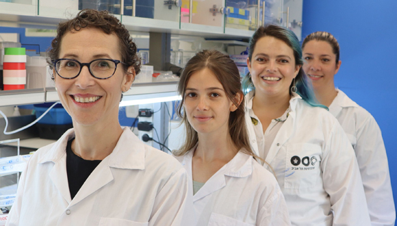
TAU research team: Dr. Inbal Ben Ami Bartal, Tamar Spectre, Estherina Trachtenberg, and Dr. Einat Bigelman (not in the photo: Keren Ruzal and Ben Kantor)

The study was led by Dr. Inbal Ben Ami Bartal of The School of Psychological Sciences and Sagol School of Neuroscience at the Tel Aviv University, in collaboration with Prof. Daniela Kaufer of the University of California and Berkeley as well as additional researchers from Stanford University and the University of Toronto. The study was published in the prestigious journal, eLife.

Can’t Multitask Anymore?

Non-invasive brain stimulation may boost mobility in the elderly and prevent falls.

Walking while simultaneously carrying out a cognitive task, like talking on a cellphone or with a companion, happens frequently throughout the day for many of us. The concurrent performance of two tasks requires the ability to split attention. For older people, difficulties performing another task while walking or standing reflect an existing and/or a potential problem concerning both functions. It also means an increased risk of falling, which can have many severe and undesirable consequences for older adults.

Tel Aviv University researchers sought to examine the benefits of very low intensity, non-invasive electrical stimulation of various parts of the brain, on the capability of older adults to walk or stand while simultaneously carrying out a cognitive task, a common dual-task situation that can determine their overall functionality. They hoped that this might improve their ability to perform both tasks simultaneously in a safer manner. The researchers found that when stimulating the dorsal lateral pre-frontal cortex (DLPFC), a cognitive brain area responsible for dividing attention and executive functions, the immediate, negative impact of a dual-task on standing and walking performance was significantly reduced.

The study team under the leadership of Prof. Jeffrey Hausdorff of the Sackler Faculty of Medicine, the Sagol School of Neuroscience, and the Tel Aviv Sourasky Medical Center (Ichilov), and Dr. Brad Manor at Harvard Medical School, as well as researchers from Harvard University, research and medical institutions in the US and Spain, and the Tel Aviv Sourasky Medical Center (Ichilov). The study was published in the Annals of Neurology, the journal of the American Neurological Association. The research was funded by a grant from the US-Israel Binational Science Foundation.

Gentle Power

The study cohort included 57 subjects over the age of 70. Each of them was tested by 4 different treatments:

  • Sham, designed not to have any influence at all, but to rule out any placebo effects;
  • Stimulation of a cognitive area of the brain (DLPFC) that is responsible for dividing attention;
  • Stimulation of a sensory-motor area of the brain which contributes to the regulation of walking;
  • Simultaneous stimulation of both areas – motor and cognitive – together. 

Each treatment included non-invasive stimulation using a very low-intensity electric current for 20 minutes. Immediately upon the conclusion of the treatment, the walking and standing sway of each subject were evaluated, with and without the request to also perform a cognitive task.

The study showed that stimulation of the cognitive area, whether alone or together with the stimulation of the motor area, reduced the negative effects of the cognitive task on walking and standing stability by about 50%.  Stimulation of the sensory-motor area alone and sham stimulation did not improve the subjects’ performance. The researchers explain that, since the stimulation is gentle, it does not activate brain neurons but only increases their excitability; in other words, it facilitates the ability of the patient to activate those neurons in his or her brain.

“In our study, we demonstrated that a low-level, gentle stimulation of a specific cognitive area of the brain can improve the performance of older adults when they carry out the double task of walking or standing in place while at the same time performing a cognitive task, at least within the immediate time range,” says Prof. Hausdorff.  

“We hope that a series of treatments will lead to similar positive results over a more protracted period: to improve standing stability and walking capability, diminish the risks of falling, and perhaps also enhance cognitive function among the elderly population. This treatment is safe, and we hope that, in time, people will be able to undergo self-treatment in their own homes. Additionally, we foresee the possibility of combining this type of therapy with exercise and other modes of intervention that can help to improve walking, to enhance thinking, and to reduce the risk of falls. There is evidence that combined therapy could prove to be the most effective solution, but further research is required to examine this,” he concludes.

Why Do We Squabble Over The AC?

New study reveals the evolutionary reason why women feel colder than men.

Why do women typically bring a sweater into work while their male counterparts feel comfortable wearing short sleeves in an air-conditioned office? Researchers at Tel Aviv University’s School of Zoology offer a new evolutionary explanation for the familiar scenario and can share that this phenomenon is not unique to humans, with many male species of endotherms (birds and mammals) preferring a cooler temperature than the females. The researchers propose that males and females feel temperature differently, explaining that this is a built-in evolutionary difference between the heat-sensing systems of the two sexes, related, among other things, to the reproduction process and caring for offspring.

The study was led by Dr. Eran Levin and Dr. Tali Magory Cohen from the School of Zoology and the Steinhardt Museum of Natural History at Tel Aviv University, Yosef Kiat from the University of Haifa, and Dr. Haggai Sharon, a pain specialist from Tel Aviv University’s Sackler Faculty of Medicine and the Tel Aviv Sourasky Tel Aviv Medical Center (Ichilov Hospital). The article was published in the Journal Global Ecology and Biogeography.

Separation During Breeding Season

The new study included an in-depth statistical and spatial analysis of the distribution of dozens of bird and bat species living in Israel, along with a comprehensive review of the international research literature on the subject.

A study of the research literature reveals several examples of a similar phenomenon observed in many species of birds and mammals. In migratory bird species, males spend the winter in colder areas than females (it should be noted that in birds, the segregation between the sexes takes place outside of the breeding season, since the males participate in the raising of the chicks).

Amongst many mammals, even in species that live in pairs or in mixed groups all their lives, the males prefer shade whereas the females prefer sunlight, or the males ascend to the peaks of mountains while the females remain in the valleys.

Following the literature review, the researchers conducted their own research. They sampled information collected in Israel over the course of nearly 40 years (1981-2018) on thousands of birds from 13 migratory bird species from 76 sites (data from Birdlife Israel and the Steinhardt Museum of Natural History) and 18 species of bats from 53 sites (data from the researchers and the Society for the Protection of Nature.) In total, the study included more than 11,000 individual birds and bats, from Mount Hermon in the north to Eilat in the south.

The reasoning behind the choice of birds and bats for the study is the fact that they fly and are therefore highly mobile, and the researchers hypothesized that the spatial separation between the sexes – sometimes extending to different climatic zones – would be particularly clear in these groups. Moreover, Israel’s significant climate diversity allowed them to study individual animals of the same species that live in very different climatic conditions.


Illustrative Photo: Bats in Cave

The findings of the study clearly demonstrated that males prefer a lower temperature than females, and that this preference leads to a separation between the sexes at certain periods during the breeding cycles, when the males and females do not need, and may even interfere, with each other.

Dr. Levin: “Our study has shown that the phenomenon is not unique to humans; among many species of birds and mammals, females prefer a warmer environment than males, and at certain times these preferences cause segregation between the two species. In light of the findings, and the fact that this is a widespread phenomenon, we have hypothesized that what we are dealing with is a difference between the females and males’ heat-sensing mechanisms, which developed over the course of evolution. This difference is similar in its essence to the known differences between the pain sensations experienced by the two sexes, and is impacted by differences in the neural mechanisms responsible for the sensation and also by hormonal differences between males and females.”

Dr. Levin, who among other things studies the physiology and behavior of bats, noted in his previous studies that during the breeding season males and females tend to segregate, with the males inhabiting cooler areas. For example, entire colonies in caves on the slopes of Mount Hermon are composed of only males during the breeding season, while in the warmer area of the Sea of Galilee there are mainly females, who give birth and raise their pups there.

Dr. Magory Cohen notes that this difference has a number of evolutionary explanations. First, the separation between males and females reduces competition over resources in the environment, and keeps away males who may be aggressive and endanger the babies. Furthermore, many female mammals must protect their offspring at a stage when they are not yet able to regulate their body temperature on their own, so they developed a preference for a relatively warm climate.

Giving Each Other Some Space

The phenomenon can also be linked to sociological phenomena observed in many animals and even in humans; in a mixed environment of females and males the females tend to have much more physical contact between themselves, whereas males maintain more distance and shy away from contact with each other.

It appears the difference in thermal sensation did not come about so we can argue with our partners over the air conditioning. Rather, we are meant to give each other some space so that each person can enjoy some peace and quiet. Question is, who gets the couch?

The Immune System’s Double Agents

The Immune System’s Double Agents

Glioblastoma is the most common type of brain cancer, and one of the most violent and deadly cancers in humans; the average life expectancy of glioblastoma patients is 12 to 15 months from the moment of detection. Usually, the scientific monitoring of the development of the cancerous tumor in animal models is carried out without an active immune system, in order to enable the absorption and growth of cancer cells in the body. The disadvantage of this commonly-used model lies in the fact that the immune system either does not exist or does not function properly, which prevents researchers from monitoring the interaction between it and the tumor cells.

A new Tel Aviv University study examined for the first time the development of a glioblastoma cancerous tumor in animal models with a normal immune system in order to best simulate the development of the tumor in humans. The findings showed that there are immune system cells that, despite the fact that their primary function is to attack and kill the cancer cells, actually act as “double agents” that increase and intensify the aggressiveness and threat of the tumor.

The study was led by Dr. Dinorah Friedmann-Morvinski of The George S. Wise Faculty of Life Sciences and Sagol School of Neuroscience, and her PhD student Prerna Magod. Also participating in the study were Dr. Liat Rousso-Noori and Ignacio Mastandrea, also from the Faculty of Life Sciences, as well as researchers from the Sackler Faculty of Medicine and the Weizmann Institute of Science. The study was published in the prestigious journal Cell Reports.

Switching Sides

In the study, the researchers found that cells called neutrophils play a critical role in interacting with the cancerous growth. Neutrophils are immune system cells that originate in the bone marrow, and whose purpose is to “swallow” or kill bacteria and fungi and fight the infections caused by them. “Neutrophils are the front-line soldiers of the immune system,” explains Dr. Friedmann-Morvinski. “When a tumor begins to develop, the neutrophils are among the first to mobilize and attack it in order to eliminate it.”

The researchers also found that the neutrophils remain near the tumor throughout its development, and are continuously and consistently recruited from the bone marrow. The surprising thing that was discovered during this study is that the neutrophils “change sides:” Whereas at first, with the onset of the initial tumor, the neutrophils fight it, over time the neutrophils recruited to the cancerous area begin to support its development.

Dr. Friedmann-Morvinski: “We learned that the neutrophils actually change their role. They are mobilized by the tumor itself, and from being anti-cancerous, become pro-cancerous; as a result, they aggravate the damage that the tumor itself creates.”

Moreover, the researchers found that the process by which the neutrophils change their properties can take place remotely, even before they progress towards the tumor itself.

“The study showed that the change in the properties of neutrophils takes place in the bone marrow itself – where there is no tumor at all: the cancerous tumor is located only in the brain, and from there it succeeds in changing the properties of the cells it recruits,” adds Dr. Friedmann-Morvinski.

Seeking to Boost Effectiveness of Immunotherapy

“The new findings of this study may also shed light on immunotherapeutic therapies, which have been gaining a lot of momentum in recent years. In one type of immunotherapy treatment, T cells [important white blood cells of the immune system] are removed from the patient’s body, processed, and returned to the body with increased healing abilities. One of the major problems today is that even these cells, that have been sent in order to heal, are suppressed and their actions stifled. If we know how to change the interaction between neutrophils and T cells so that they are not suppressed, this will have implications for the effectiveness of immunotherapy.”

These revelations may be the first step towards deciphering the mechanism of interaction between the immune system and cancerous tumors.

Using ‘Good’ Bacteria to Fight ‘Bad’ Bacteria

Antibiotic resistance is an ever-growing worry to the medical profession, with the World Health Organization recently defining it as one of the most significant dangers to public health and food safety. A new technology from Tel Aviv University will make it possible to insert ‘good’ bacteria into the body, or into diverse environmental niches, for the purpose of injecting toxins and eliminating ‘bad’ bacteria. The breakthrough technology can be suited to target different kinds of bacteria and may become a biological replacement for antibiotics, potentially saving many lives.

The study was conducted by Dr. Dor Salomon, Dr. Biswanath Jana and Kinga Kappel of the Department of Clinical Microbiology and Immunology at the Sackler Faculty of Medicine.

Taking Advantage of Killing System

Beneficial bacteria and pathogenic bacteria, bacteria that can cause disease, have fought each other over resources and nutrients since the dawn of time, and have developed a variety of sophisticated mechanisms that neutralizes their competition. Understanding the mechanisms that mediate these bacterial wars will enable their utilization and conversion into new tools that will be used to treat diseases caused by antibiotic-resistant bacteria.

TAU researchers built a system which allows them to engineer ‘good’ bacteria that can recognize disease-causing bacteria, attack the latter exclusively with toxins and neutralize them. Dr. Dor Salomon explains, “We know how to change and control every component in the system and create a bacterium that neutralizes different strains of bacteria. This is proof of feasibility, showing that we have the knowledge and ability to create bacteria that take advantage of this killing system and may serve as antibiotic treatments. Such bacteria could replace the classic antibiotics that we currently use in a variety of scenarios”.

High Degree of Control

The researchers ‘borrowed’ a toxin injection system – known as a Type 6 Secretion System – from a pathogenic bacterium and introduced it into a ‘friendly’ bacterium, Vibrio natriegens. This ‘friendly’ bacterium is not harmful to humans or animals and can survive and reproduce under a variety of conditions. The injection system is similar to a poisoned arrow shot from a bacterium towards neighboring bacteria.

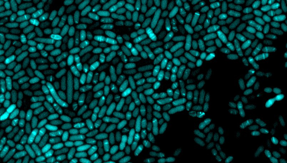
Photo: Vibrio bacteria expressing a structural component of the type 6 secretion system fused to a green fluorescent protein, allowing us to visualize the assembly of the secretion 

Toxins carried on the arrow then mediate the elimination of competing bacteria. With the help of central regulator protein that they identified, researchers were able to produce an ‘operating switch’ for the system and cause it to ‘turn on’ only in response to recognizing desirable environmental conditions. In addition, researchers proved it possible to control the type and amount of toxins that are loaded on the arrow, thus adjusting the system’s killing range.

Serving Many Purposes

The system in its current form is predominantly suited for preventing and treating bacterial infections that affect the production of food from marine animals. It can then be adapted to treat pathogenic bacteria in humans, farm animals, plants and the environment.

“Ramot The Technology Transfer Company of Tel Aviv University has filed a patent application to protect the technology and its application. Many companies in the field have already expressed interest in this sophisticated system developed by Dr. Dor Salomon, Dr. Biswanath Jana and Kinga Kappel”, says Keren Primor Cohen, CEO, Ramot at TAU. The study was published in the peer-reviewed EMBO Reports.

Recruiting ‘Fighting Cells’ to Destroy Tumors

TAU discovery of cell interaction may serve as basis for improved cancer treatments.

Tel Aviv University researchers have found that our body’s natural defenses can help destroy malignant tumors. The team found that white blood cells called ‘eosinophils’ fight cancer in two ways: they can destroy the cancer cells themselves, and also recruit the immune system’s cancer-fighting T-cells – another type of white blood cells key to protecting the body against infection. These findings may contribute to the development of new immunotherapies.

Fighting Cancer from Within

The discovery comes as the rising number of cancer cases every year has contributed to a boom in immunotherapy, a treatment that activates the body’s immune system to fight disease. Compared to traditional techniques like chemotherapy, immunotherapy generally leads to longer protection from cancer and fewer side effects.

“Enhancing the number and power of T-cells is one of the main targets of immunotherapy treatments administered to cancer patients today,” said lead researcher, Prof. Ariel Munitz of TAU’s Department of Microbiology and Clinical Immunology at the Sackler Faculty of Medicine. “We discovered a new interaction that summons large quantities of T-cells to cancer tissues, and our findings may have therapeutic implications. Ultimately, our study may serve as a basis for the development of improved immunotherapeutic medications that employ eosinophils to fight cancer.”

Repurposing Cells’ “Destructive” Qualities

Eosinophils produce powerful destructive proteins originally intended for fighting parasites. However, in the modern Western world, where high levels of hygiene have significantly reduced the risk of many parasites, eosinophils often have a negative impact on humans, inducing phenomena like allergies and asthma.

Considering the destructive power of eosinophils, the researchers decided to test the potential benefits of these white blood cells if turned against cancer cells.

For two main reasons, they decided to focus their study on lung metastases, or cancer that started in another part of the body and spread to the lungs: “First, metastases, and not the primary tumors, are often the main problem in treating cancer. The lungs are a major target for the metastasis of many types of cancer,” said Prof. Munitz. “Second, in a preliminary study we demonstrated that eosinophils gather in tumors developing in mucous tissues like the lungs, and therefore assumed that they would be found in lung metastases as well.”

Summoning Reinforcement

The researchers examined tissue samples taken from breast cancer patients. They found that the eosinophils reach the lungs and penetrate the cancer tissues, where they often release their destructive proteins and summon T-cells for reinforcement. Ultimately, T-cells gather in the affected lungs, slowing the growth of tumors.

Additionally, the researchers found that in the absence of eosinophils, the lung tumors were much larger than those exposed to the white blood cells. These findings led to the conclusion that eosinophils fight cancer effectively.

Along with Prof. Munitz, the study was led by TAU PhD student Sharon Grisaru. The findings were published in the journal Cancer Research, published by the American Association for Cancer Research. The initiative was funded by ICRF (Israel Cancer Research Fund), the Israel Cancer Association, ISF (the Israel Science Foundation) BSF (U.S.-Israel Binational Science Foundation) and GSK.  

Featured image: Illustration: Eosinophil, a white blood cell in 3D

TAU Team Reverses Early Signs of Alzheimer’s

New non-drug method holds promise for preventative therapies.

Approximately 50 million people worldwide live with Alzheimer’s or other related forms of dementia. Alzheimer’s disease leads to memory loss and impairment in cognitive function, and is the most common cause of dementia among older adults. While certain treatments can help reduce symptoms and sometimes reduce disease progression, there is currently no way to prevent or cure Alzheimer’s.

Amid that backdrop, researchers from Tel Aviv University have developed a process for reversing the precursors of the disease, providing a promising foundation for new preventative therapies. This marks the first time that a non-drug therapy has proven effective in preventing the core biological processes that lead to the development of Alzheimer’s, providing hope that we will now be able to fight one of the greatest challenges to the Western world.

Targeting the Root of Alzheimer’s

Using hyperbaric oxygen therapy (HBOT), in which subjects breathe 100% oxygen in a special chamber of high atmospheric pressure, the researchers were able to reverse brain damages associated with the biological hallmarks of Alzheimer’s.

 “By treating the root problem that causes cognitive deterioration with age, we are in fact mapping out the way to prevention,” says co-lead researcher Prof. Shai Efrati.

Often used to treat carbon monoxide poisoning and infections that starve tissues of oxygen, hyperbaric therapy, when applied in a specific way, has previously been found capable of repairing damaged brain tissue and renewing growth of blood vessels and nerve cells in the brain. Therefore, the researchers tested its potential for Alzheimer’s.

“After a series of hyperbaric treatments, elderly patients who were already suffering from memory loss showed an improvement of blood flow to the brain as well as a real improvement in cognitive performance,” said co-lead investigator Prof. Uri Ashery.

The new approach devised by the researchers unequivocally improved characteristics commonly associated with Alzheimer’s disease. Specifically, the hyperbaric treatment resulted in:

  • Improved memory in 16.5% of patients on average
  • Increased blood flow in 16%-23% of cases
  • Improved attention and concentration in 6% of patients
  • Improved information processing speed in 10.3% of all cases

A Future Without Alzheimer’s?

 “Our findings provide hope that we will now be able to fight one of the greatest challenges to the Western world. According to our findings, hyperbaric therapy given at a young age is likely to prevent this severe disease entirely,” explains TAU team member Dr. Ronit Shapira.

The approach was first tested in laboratory settings followed by testing in patients over the age of 65 in stages of deteriorating mental function that often precede Alzheimer’s and dementia. The therapy included a series of 60 treatments in hyperbaric chambers over a period of 90 days.

The study is part of a comprehensive research program focused on reversing processes of aging and its accompanying ailments. The researchers note that the findings are an encouraging step toward new approaches to preventing Alzheimer’s by addressing not only the symptoms or targeting biomarkers, but the core pathology and biology responsible for the disease’s development.

The Tel Aviv University team that led the study included Prof. Shai Efrati of the Sackler Faculty of Medicine and the Sagol School of Neuroscience, Prof. Uri Ashery and  Dr. Pablo Blinder of the The George S. Wise Faculty of Life Sciences and the Sagol School of Neuroscience, and Dr. Ronit Shapira and Dr. Amir Hadanny. They are all affiliated with the Shamir Medical Center. The findings were published in the journal Aging.

Featured image: Alzheimer’s Disease on MRI

Nicotine Testing of Children Curbs Parents’ Smoking

70% of children of smokers suffer from secondhand smoking.

Warnings against the dangers of smoking can be read on every cigarette box and in every advertisement for smoking brands. Those who smoke endanger themselves and also those around them, who inevitably become passive smokers. This way, parents who smoke harm the health of their own children. A first-of-its-kind study in Israel by researchers from the Sackler Medical School of Tel Aviv University uncovers alarming data about secondhand smoking by children of smokers: According to the study, nicotine residues were found in the hair samples of 7 out of 10 children who participated. The research team found that parental behavior may be changed through regular monitoring of children’s exposure.

Nicotine Residue in Children’s Hair

The study was conducted under the leadership of a team of experts from the Tel Aviv University School of Medicine headed by Prof. Leah (Laura) Rosen of the School of Public Health together with researchers Dr. Vicki Myers, Prof. Nurit Guttman, Ms. Nili Brown, Prof. Mati Berkovitch, and Dr. Michal Bitan. Prof. David Zucker of the Hebrew University of Jerusalem and Dr. Anna Rule of Johns Hopkins University in the US also participated in the study. The study was published in the prestigious journal, Nicotine & Tobacco Research.

In the study, the researchers sought to examine whether raising awareness of children’s exposure by providing objective feedback might change the parents’ behavior and child exposure. 140 Israeli families participated in the study, parents of children up to age 8, at least one parent being a smoker. The smoking average per household was 15 cigarettes per day, where one third of the respondents reported that they smoke inside the home, and one third said that they smoke on the terrace but not inside the home.

First, researchers tested children’s level of exposure via a biomarker, nicotine in hair, which indicates cumulative exposure to tobacco smoke. The researchers took hair samples from the children and tested the nicotine levels in each sample (it is important to note that the test was for nicotine that became an integral part of the strand of hair and not just outside precipitate.) The findings were very concerning: Nicotine residue was found in the hair of 70% of the children tested. Only 29.7% of the children tested did not show nicotine residue in their hair samples. 

The researchers divided the families into two groups: one group underwent comprehensive instruction about the effects and dangers of exposure to smoking, including feedback and information about the test results. The group was also given tools to protect their children from exposure to cigarette smoke and a recommendation to keep their home and car smoke-free. The second group received feedback about nicotine levels in the children’s hair after six months, at the end of the study.

Limiting Children’s Exposure

Six months after the start of the study, the researchers conducted additional nicotine tests on the children’s hair, and one could already see a significant improvement in the data: Among the group that received comprehensive training, the percentage of children whose hair samples contained nicotine decreased from 66% to 53%, whereas in the second group (which did not receive training at the start of the study), the percentage of children whose hair samples contained nicotine decreased from 74% to 49%. Thus, just testing the children, without even informing families of the results, was enough to seemingly change parents’ behaviors.

The researchers theorize that the knowledge that the children were tested for tobacco smoke exposure, and that additional testing was planned at six months, resulted in the parents changing their behavior and reducing the children’s exposure. As a result of the study’s findings, the researchers recommend considering conducting such testing to measure exposure on a routine basis among young children in Israel.

The Right to Breathe Smoke-Free Air

Prof. Leah Rosen: “To our great dismay, according to the Ministry of Health’s data, approximately 60% of small children in Israel are exposed to secondhand smoke and its harmful effects. Based on the study’s findings, we believe that conducting nicotine testing – in the hair, urine, or using other testing methods – for every young child in Israel, may change parents’ perceptions about exposing their children to tobacco smoke. Changing this perception can also result in changing behavior, exposure levels, and even social norms regarding passive exposure to smoking – both exposure of children as well as exposure of adults.”

“We call upon smokers to avoid smoking anyplace where non-smokers and in particular, at-risk populations, including children, pregnant women, elderly, and those who are ill, could be exposed. Non-smokers must understand that there is genuine risk in exposure to tobacco smoke, and they must insist upon their right and the right of their children and family members to breathe air that is smoke-free everywhere. Of course, the government has a central role in enforcing laws pertaining to smoking in public places and continuing to enact laws to protect the individual everywhere from exposure to secondhand smoke.” 

Victoria

Phone: +61 3 9296 2065
Email: office@aftau.asn.au

New South Wales

Phone: +61 02 7241 8711
Email: davidsolomon@aftau.org.au

Western Australia

Phone: :+61 411 223 550
Email: clivedonner@thelinqgroup.com