Tag: Medicine

TAU Researcher Fights Epidemics Both Viral and Virtual

Dan Yamin can detect any kind of contagious outbreak

TAU’s Dr. Dan Yamin has developed a data tracking system applicable both to infectious diseases like coronavirus and to anti-Israel bias on social media. He cites human behavior as a key factor in the transmission of both. Yamin, who heads the Lab for Epidemic Modeling and Analysis at TAU’s Fleischman Faculty of Engineering, says that his approach is based on what traditional epidemiology lacks – data on human behavior. “At the core of any transmission process lies contact mixing patterns,” explains Yamin. “These patterns represent the social interactions of individuals,” adding that, when it comes to the spread of diseases, “whoever doesn’t consider these elements misses the point completely”. Together with Prof. Irad Ben-Gal, head of TAU’s Laboratory of AI, Machine Learning, Business & Data Analytics (LAMBDA), Yamin developed a tool for predicting transmission dynamics based on people’s movements tracked on their mobile phones. When COVID-19 broke out in Israel, Yamin consulted for Israel’s Health Ministry, predicting local outbreaks with this phone data system. “The tool is not only helpful for local detection of the virus but also for creating simulations of the virus’ spread, telling us what will happen if one policy is replaced with another,” he says. For example, Yamin’s team recommended to the Health Ministry that daycare centers should re-open, based on data they collected. Additionally, Yamin found that targeted lockdowns for high-risk groups and localized infection clusters are up to 5 times more efficient in reducing mortality as opposed to a nationwide lockdown strategy. This finding led the Israeli government to adopt its current targeted lockdown approaches. Now, months later, Yamin and his team are developing a tool for early detection of COVID-19 infection based on mobile phone sensors which measure step counts, sleeping habits and other parameters.

Think viral, tweet viral

Before joining TAU, Yamin completed a post-doctoral fellowship at Yale University’s School of Public Health. While there, he was disturbed by the level of anti-Israel sentiment on American social networks and its ability to go viral. He immediately made the connection. No paragraph breakBased on the same patterns he studied in disease transmission, Yamin began creating a system that uses artificial intelligence to identify how certain groups use viral marketing tactics to spread anti-Semitic and anti-Israel messages. The system, known as Iron Dome for Social Media, aims to track and identify malicious content with potential to go viral in social media terms. Yamin explains that people who retweet posts casually are much like asymptomatic disease carriers. Many Twitter users will pass on information with covert or explicit anti-Semitic messages unintentionally. Choosing when to respond on social media is a delicate matter. Hence, Yamin suggests using AI, such as in his Iron Dome system, to assist with the decision-making process. “Being proactively pro-Israel on social media is not always the best approach,” says Dr. Yamin. “Most anti-Israel tweets are not viral, so why waste time on tweets that won’t go anywhere?”

The next generation of disease control

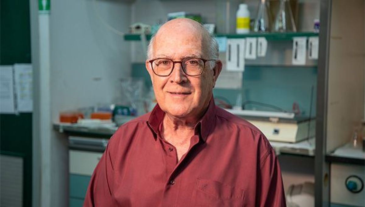
As Israel and the world face the second wave of COVID-19, Yamin says, “for the time being, we need to live with this virus. If we act responsibly and maintain the daily routine for the vast majority of the population, we will not reach catastrophe”. Looking ahead, Yamin believes data-based methodologies like his are crucial in managing future viral diseases. As such, Yamin will be a key member of TAU’s multidisciplinary Center for Combating Pandemics, the first center of its kind in the world. “Data systems such as this one can substantially improve the accuracy of medical diagnosis in the future,” he says. Dr. Dan Yamin (Photo: Moshe Bedarshi)

TAU study: Oxygen therapy improves cognitive function in seniors

Research Published in Aging first to Show Enhanced Brain Function and Cognitive Capabilities Resulting from Novel Therapy

The Sagol Center for Hyperbaric Medicine and Research at Shamir Medical Center, together with Sackler School of Medicine and Sagol School of Neuroscience at Tel Aviv University announced that, for the first time, in humans, a peer-reviewed study has demonstrated that hyperbaric oxygen therapy (HBOT) can significantly enhance the cognitive performance of healthy older adults. The main areas of improvement were attention, information processing speed, executive function, in addition to the global cognitive function, all of which typically decline with age. Moreover, there was a significant correlation between the cognitive changes and improved cerebral blood flow in specific brain locations. The study was published on July 15th, 2020 in the peer reviewed journal Aging, entitled: Cognitive enhancement of healthy older adults using hyperbaric oxygen: a randomized controlled trial. Professor Shai Efrati, Head of the Sagol Center for Hyperbaric Medicine and Research, and Head of Research & Development at Shamir Medical Center, and an Associate Professor at Sackler School of Medicine and Sagol School of Neuroscience at Tel Aviv University, and Dr. Amir Hadanny, the Sagol Center for Hyperbaric Medicine and Research, designed the study based on a unique HBOT protocol developed at the Sagol Center over the past 10 years. The randomized controlled clinical trial included 63 healthy adults (>64) who underwent either HBOT (n=33) or a control period (n=30) for three months. The study’s primary endpoint included a change in general cognitive function measured by a standardized comprehensive battery of computerized cognitive assessments before and after the intervention or control. Cerebral blood flow (CBF) was evaluated by a novel magnetic resonance imaging technique for brain perfusion. “Age-related cognitive and functional decline has become a significant concern in the Western world. Major research efforts around the world are focused on improving the cognitive performance of the so-called ‘normal’ aging population,” said Prof. Efrati. “In our study, for the first time in humans, we have found an effective and safe medical intervention that can address this unwanted consequence of our age-related deterioration.” “Over years of research, we have developed an advanced understanding of HBOT’s ability to restore brain function. In the past, we have demonstrated HBOT’s potential to improve/treat brain injuries such as stroke, traumatic brain injury and anoxic brain injury (due to sustained lack of oxygen supply) by increasing brain blood flow and metabolism,” explained Dr. Amir Hadanny. “This landmark research could have a far-reaching impact on the way we view the aging process and the ability to treat its symptoms.” During HBOT, the patient breaths in pure oxygen in a pressurized chamber where the air pressure is increased to twice that of normal air. This process increases oxygen solubility in the blood that travels throughout the body. The added oxygen stimulates the release of growth factors and stem cells, which promote healing. HBOT has been applied worldwide mostly to treat chronic non-healing wounds. There is a growing body of evidence on the regenerative effects of HBOT. The researchers have demonstrated that the combined action of delivering high levels of oxygen (hyperoxia) and pressure (hyperbaric environment), leads to significant improvement in tissue oxygenation while targeting both oxygen and pressure sensitive genes, resulting in restored and enhanced tissue metabolism. Moreover, these targeted genes induce stem cell proliferation, reduce inflammation and induce generation of new blood vessels and tissue repair mechanisms. “The occlusion of small blood vessels, similar to the occlusions which may develop in the pipes of an ‘aging’ home, is a dominant element in the human aging process. This led us to speculate that HBOT may affect brain performance of the aging population,” Prof. Efrati explained. “We found that HBOT induced a significant increase in brain blood flow, which correlated with cognitive improvement, confirming our theory. One can conjecture that similar beneficial effect of HBOT can be induced in other organs of the aging body. These will be investigated in our upcoming research.” The research group leader, Professor Shai Efrati, who serves as director of The Sagol Center for Hyperbaric Medicine and Research, and is an Associate Professor at Sackler School of Medicine and Sagol School of Neuroscience at Tel Aviv University, also disclosed his role with Aviv Scientific LTD, which has developed a comprehensive program that includes HBOT treatment, cognitive and physical training and nutritional coaching, to enhance brain and body performance of aging adults based on the Sagol HBOT protocol at Aviv Clinics. Prof. Efrati serves as Chair of Aviv Scientific’s Medical Advisory Board

TAU-led Team Destroys Cancer Cells with Ultrasound

Breakthrough method may be applicable to Parkinson’s, Alzheimer’s and more

An international research team, headed by Dr. Tali Ilovitsh from the Biomedical Engineering Department at Tel Aviv University, developed a noninvasive technology platform for gene delivery into cancer cells (breast cancer). The technique combines ultrasound together with tumor-targeted microbubbles. Once the ultrasound is activated, the microbubbles explode like smart and targeted warheads, creating holes in cancer cells’ membranes, and enabling the gene delivery. The two-year research was recently published in the prestigious journal Proceedings of the National Academy of Sciences (PNAS).

Dr. Ilovitsh developed this breakthrough technology during her post doctorate research at the lab of Prof. Katherine Ferrara at Stanford University. The technique utilizes low frequency ultrasound (250 kHz) to detonate microscopic tumor-targeted bubbles. In vivo, cell destruction reached 80% of tumor cells.

Dr. Ilovitsh explains: “Microbubbles are microscopic bubbles filled with gas, with a diameter as small as one tenth of a blood vessel. At certain frequencies and pressures, sound waves cause the microbubbles to act like balloons: they expand and contract periodically. This process increases the transfer of substances from the blood vessels into the surrounding tissue. We discovered that using lower frequencies than those applied previously, microbubbles can significantly expand, until they explode violently. We realized that this discovery could be used as a platform for cancer treatment and started to inject microbubbles into tumors directly.”

Dr. Ilovitsh and the rest of the team used tumor-targeted microbubbles, that were attached to tumor cells’ membranes at the moment of the explosion, and injected them directly into tumors in a mouse model. “About 80% of tumor cells were destroyed in the explosion, which was positive on its own,” says Dr. Ilovitsh. “The targeted treatment, which is safe and cost effective, was able to destroy most of the tumor. However, it is not enough. In order to prevent the remaining cancer cells to spread, we needed to destroy all of the tumor cells. That is why we injected an immunotherapy gene alongside the microbubbles, which acts as a Trojan horse, and signaled the immune system to attack the cancer cell.”

On its own, the gene cannot enter into the cancer cells. However, this gene aimed to enhance the immune system was co-injected together with the microbubbles. Membrane pores were formed in the remaining 20% of the cancer cells that survived the initial explosion, allowing the entry of the gene into the cells. This triggered an immune response that destroyed the cancer cell.

“The majority of cancer cells were destroyed by the explosion, and the remaining cells consumed the immunotherapy gene through the holes that were created in their membranes. The gene caused the cells to produce a substance that triggered the immune system to attack the cancer cell. In fact, our mice had tumors on both sides of their bodies. Despite the fact that we conducted the treatment only on one side, the immune system attacked the distant side as well.”

Potential treatment for brain diseases such as Parkinson’s and Alzheimer’s

Dr. Ilovitsh says that in the future she intends to attempt using this technology as a noninvasive treatment for brain related diseases such as brain tumors and other neurodegenerative conditions such as Alzheimer’s and Parkinson’s disease. “The Blood-Brain barrier does not allow for medications to penetrate through, but microbubbles can temporarily open the barrier, enabling the arrival of the treatment to the target area without the need for an invasive surgical intervention.”

Photo: Dr. Tali Ilovitsh

New nanomedicines for mRNA therapeutics in breast cancer and heart failure

The project has been awarded a total of 14.9 million EUR; Prof. Dan Peer is leading the mRNA targeting work package.

TAU researcher Prof. Dan Peer, from the school of Molecular Cell Biology and Biotechnology, is one of 11 partners in the international project EXPERT that has been awarded a total of 14.9 million EUR from the EU Horizon 2020. The project is working to find efficient ways to deliver protein coding mRNA by using various nanoparticles for the treatment of breast cancer and myocardial infarction, which are two of the most pressing health challenges in European society today. Prof. Dan Peer, Director, Laboratory of Precision NanoMedicine, School of Molecular Cell Biology and Biotechnology, George S. Wise Faculty of Life Sciences and Department of Materials Sciences and Engineering, Iby and Aladar Fleischman Faculty of Engineering, Tel Aviv University Center for Nanoscience and Nanotechnology and Tel Aviv University Cancer Biology Research Center.

Dan, What is the research about in the EXPERT project?

“It is about developing mRNA therapy for the treatment of breast cancer. Much of it involves testing different methods to improve the delivery of mRNA to cells in vivo. These methods are fundamentally based either on lipid nanoparticles (LNPs), biological nanoparticles called exosomes, or cell penetrating peptides (CPPs). In addition to this, we intend to analyze what these nanoparticles bind to in biological fluids in order to better understand what drives uptake in specific cells types”.

What is your and TAU’s part in the project?

“Our lab was the first to show systemic, cell specific delivery of mRNA molecules that express therapeutic proteins in designated cells. We will further develop our ASSET platform for cell specific targeting of lipid nanoparticles to achieve improved delivery of therapeutic mRNAs and optimize formulations that enable systemic administration in different preclinical models. Part of the work will also consist of understanding how nanoparticle surfaces bind to host factors in blood and how this can affect the uptake of nanoparticles”.

What happens now, what is the next step?

“We will now see how these delivery methods work side by side in cell culture and animal models. The hope is then to be able to deliver an mRNA cocktail with one of the aforementioned vectors for the treatment of triple-negative breast cancer. In parallel, these vectors will also be evaluated for delivery of VEGF mRNA in the treatment of myocardial infarction”.

Tel Aviv University presents an analysis of the reaction of human antibodies to the coronavirus

Patients with severe COVID-19 develop antibodies faster than those with a mild case of the disease.

A team of researchers from Tel Aviv University and the Sharon Hospital at the Rabin Medical Center, led by Prof. Motti Gerlic and Prof. Ariel Munitz of the Department of Microbiology and Clinical Immunology at TAU’s Sackler School of Medicine, applied an innovative antibody test to about 70 COVID-19 patients at the Sharon Hospital. The researchers examined the development of antibodies targeting two different viral proteins in the patients’ bodies, and found that severely ill patients developed the antibodies at a faster rate than those with a mild case of the disease. In addition, antibodies of the type IgG were maintained in the blood of most patients throughout the study. This project has important implications for our understanding of the immune response to SARS-CoV-2, as well as future tracking of the effectiveness of vaccines and population surveys (serological tests).

A diagnostic tool

The researchers found that antibodies of the type IgM, that usually develop at the early stages of viral contagions, developed early in this case only against the protein RBD – the site at which the virus SARS-CoV-2 binds to human cells, and not against the virus’s nuclear protein. “We sampled the antibodies of about 70 COVID-19 patients at the Sharon Hospital, throughout the outbreak of the disease in Israel,” says Prof. Munitz. “Our first finding was that not all viral proteins generate a rapid immune response, but that antibodies targeting the RBD protein did develop very quickly once the symptoms appeared. This finding is quite significant, because it suggests that the test we used may be utilized as a diagnostic tool at different stages of the illness.” “The second thing we noticed, which is even more interesting, is that patients defined as severely ill developed antibodies at a faster rate than mildly ill patients, but ultimately all patients exhibited a similar immune response,” recounts Prof. Munitz. “Patients with mild, moderate and severe COVID-19 all developed the same level of antibodies. This is important, because one might have thought that the severely ill became so sick because they did not develop a sufficient amount of antibodies, and were thus unable to combat the virus effectively. We assume that the fast development of antibodies in these patients indicates that their immune system is hyper-active, but this hypothesis requires further research.”

Immunological memory

“We measured the levels of antibodies in the patients’ blood when they arrived at the hospital, during the period of hospitalization and after their release,” explains Prof. Gerlic. “We tried to understand whether the level of antibodies in their blood corresponded in any way to the severity of the illness, whether the antibodies developed in a similar way in all patients, and whether they remained in the blood for long periods of time – a critical factor for the ‘herd immunity’ we all wish to attain. We found that at later stages of the disease, about 50 days after the initial appearance of symptoms, a significant decline occurred in the presence of antibodies types IgM and IgA, regardless of the severity of the illness. In IgG-type antibodies, however, we observed only a slight decrease, even in mildly ill patients. IgG-type antibodies play an extremely important role in the immune response because they can neutralize the protein that binds the virus to human cells to enable contagion – thereby preventing the virus from penetrating the cells. We have not yet examined how the antibody actually works, and we do not know whether or not it neutralizes the virus, but the facts that these antibodies are quickly produced in all patients, and stay in the blood for a long time, suggest that they provide some level of immunity. So far, we have found that IgG-type antibodies remain in the body for two months. We will continue to monitor the patients for another year, to find out how long the antibodies remain in their bodies – hoping for the formation of an immunological memory.” In the new study the researchers from TAU used a new serological test developed in their laboratory. The IDF’s Medical Corps has already used the serological test developed by Prof. Gerlic and Prof. Munitz to detect COVID-19 antibodies in the blood of IDF soldiers. Within the next few weeks the test will be sent to the Israel’s Ministry of Health for validation, so that it may be used in population surveys. “Alongside the interesting findings,” says Prof. Munitz, “we wanted to demonstrate that our method is valid and more effective than the prevalent test for antibodies targeting viral proteins. To this end we examined samples of antibodies from the blood of COVID-19 patients, alongside samples from 200 healthy participants, taken before November 2019. We proved that our test, based on the antibodies, was able to distinguish between those who were ill and those who were not – at very high levels of sensitivity and specificity. One reason for this success is that we screen for three different antibodies: IgM that appears early and declines early, IgA – found on mucous surfaces like the lungs, and IgG, which we intend to test in the long run, because it may possibly lead to immunity.

3D printed heart used to test life-saving drugs

Pharmaceutical company Bayer will test new drugs using human heart tissues 3D-printed in Tel Aviv University

Last April, Prof. Tal Dvir of the George S. Wise Faculty of Life Sciences, the Iby and Aladar Fleischman Faculty of Engineering and his team successfully produced the first-ever 3D-printed heart, from tissue extracted from a patient. The researchers estimate that it will be possible to print personalized organs and tissues within 10-15 years, thus eliminating the need for organ donations and the risk of transplant rejection. Meanwhile, this innovative technology already has the potential to revolutionize a different medical field: drug screening.

Saving precious time

“In a Petri dish, all the cells line up in 2D, and it’s only one type of cell” says Prof. Dvir. “In contrast, our engineered tissues are 3D-printed, and therefore better resembles real heart tissues. Our printed tissues contain cardiac muscle, blood vessels and the extracellular matrix which connects the different cells biochemically, mechanically and electrically. Moving away from Petri dishes to 3D printed tissues could significantly improve drug tests, saving precious time and money with the hope of producing safer and more effective medication”. Ramot at Tel Aviv University has signed a collaboration agreement with Bayer to develop and validate a platform for in vitro cardiotoxicity screening, using human heart tissues 3D-printed in Prof. Tal Dvir’s Laboratory for Tissue Engineering and Regenerative Medicine. In upcoming years, Prof. Dvir’s team and Bayer plan to test new medication for toxicity and efficacy using printed whole human hearts. Drug candidates go through several phases of screening before reaching pharmacies. First, the new chemical compound is tested on human tissue cultures. Then, it is administered to lab animals. Finally, the drug is approved for human clinical trials. Prof. Dvir’s 3D-printed tissues could enable faster, cheaper and more efficient screening than Petri dishes. Prof. Dvir hopes to offer Bayer, in the near future, pre-clinical trials on complete printed organs. “Our agreement is just the beginning,” says Prof. Dvir. “Our end goal is to engineer whole human hearts, including all the different chambers, valves, arteries and veins – the best analogue of this complex organ – for an even better toxicological screening process.” To make further use of the application, Ramot at Tel Aviv University licensed the technology to a spin-off company called Matricelf, which first focuses on engineering personalized spinal cord implants to treat paralyzed patients. Matricelf has recently secured a large investment, allowing it to reach clinical settings in the near future.

New, innovative drugs

Keren Primor Cohen, Ramot CEO said: “Prof. Dvir’s platform groundbreaking innovation is very promising. We believe that this collaboration with Bayer will support the evaluation and development of new drugs and is a step in building long-term relations with Bayer that we hope will benefit both partners and ultimately patients.” “We are excited to start this new collaboration with Tel Aviv University, which will address a new area of early assessment of safety and tolerability of drug candidates,” said Eckhard von Keutz, Head of Translational Sciences at Bayer. “We already have a global network of partners and this new project will enable Bayer to expand its open innovation activities to Israel, which provides a dynamic ecosystem for innovation in biotech and medical research

Did climate change cause infections 6,000 years ago?

New study of human skulls finds infections peaked due to high population density, poor hygiene and climate conditions

Researchers at Tel Aviv University have discovered evidence of ear infections in the skull remains of humans living in the Levant some 15,000 years ago. “Our research seeks to determine the impact of our environment on illnesses in different periods,” says lead author Dr. Hila May of the Department of Anatomy and Anthropology at TAU’s Sackler Faculty of Medicine and the Dan David Center for Human Evolution and Biohistory Research at the Faculty of Medicine, located at the Steinhardt Museum of Natural History. “Using advanced technologies and unique methods developed in our lab, we have been able to detect signs of prolonged inflammation in the middle ear.” The researchers found a decline in morbidity as a result of ear infections following the transition from hunting and gathering to farming on account of changes in living conditions. A peak in morbidity, however, was observed in a sedentary population living about 6,000 years ago (Chalcolithic period). Dr. May says the reason for this is twofold: social and environmental: “We know from archaeological excavations of this period, similar to preceding periods, people lived in a communal area where all activities, from cooking to raising livestock, took place. As a result, the population density in the ‘home’ was high, hygiene was poor and they suffered from indoor air pollution. Two other factors known about this period – dietary change, the advent of dairy consumption, and climate change, a dip in temperature and a rise in rainfall, also contributed to the prevalence  of ear infections.”

A story in the skulls

Until the advent of antibiotics in the 20th century, ear infections developed into chronic conditions, or, due to complications, caused permanent loss of hearing or even death. “Ear infections are still a very common childhood ailment, with over 50 percent of young children today still suffering from an ear infection at one point or another,” explains Dr. May. “The reason for this is that the tubes that channel fluid from the middle ear to the mouth are underdeveloped in young children, so fluids that accumulate in the ear ultimately cause inflammation.” “A prolonged ear infection would cause permanent damage to the bony wall of the middle ear, which is remarkably preserved into adulthood, so when we sought to investigate changes in communal health over time in our region, we chose to focus on ear infections, developing a special method for doing so,” she adds. The scientists used a videoscope, a tiny camera mounted at the end of a flexible tube, which they inserted through the ear canal to the middle ear to observe its bony walls. In addition, they scanned skull remains with a high-resolution micro-CT, and also examined the middle ear’s bony wall using a light microscope.

More room, fewer infections

As living conditions improved, morbidity as a result of ear infections dropped, according to the study. “Houses were larger and featured several rooms, including separate areas for specific activities, i.e. the kitchen was set up in a separate room or outside, and livestock were kept in a separate area,” she says. “The change in lifestyle and climate is reflected in a decline in morbidity.” “Our study deals with the impact of the environment and social behavior on morbidity rates, and to do so, we examined a common disease that has accompanied humanity since inception – the ear infection,” concludes Dr. May. “Understanding how diseases appear, spread and disappear throughout human history can help prevent and find solutions to contemporary illnesses. The study clearly points out risk factors and shows how lifestyle changes can affect the incidence of the disease. In both ear infections and COVID-19, social distancing and adherence to hygiene reduced the spread of infection, while close quarters and unhygienic living conditions saw infections spike.”

New TAU study tracks coronavirus spread patterns in Israel

Research finds approximately 70% of the infections in Israel were caused by a SARS-CoV2 strain imported from the United States

A  team of Tel Aviv University researchers led by Dr. Adi Stern of the School of Molecular Cell Biology and Biotechnology at TAU’s George S. Wise Faculty of Life Sciences have conducted the first large-scale genomic sequencing of the novel coronavirus strain that has infected to date over 16,500 people in Israel. The scientists harnessed their genomic map to pinpoint mutations indicating where the virus originated from and later spread to within Israel. The study is based on an analysis of the genomic sequences of over 200 patients at hospitals across Israel, who together constitute a representative sample of the general population. TAU doctoral students Daniel Miller, Noam Harel, Talia Kostin, Omer Tirosh and Moran Meir conducted the research for the study in collaboration with scientists at Emory University, Gertner Institute, Chaim Sheba Medical Center, the Holon Institute of Technology, Assuta Hospital Ashdod, Hadassah Ein Karem Medical Center, Soroka Medical Center, Barzilai Medical Center, Poriya Medical Center, and the Genome Center at the Technion Institute of Technology.

The origins of coronavirus

“The novel coronavirus is characterized by mutations that occur at a set pace,” explains Dr. Stern. “These mutations do not affect the virus, i.e. it remains stable, but these mutations can help us trace the chain of infection from country to country. After the pandemic broke out in Wuhan, for example, one or two mutations occurred, and one virus with a mutation may have migrated to Europe where it experienced additional mutations, and from there it traveled to the United States, and so on. “We can look at these mutations as a kind of barcode that helps us keep track of the progression and transformation of the coronavirus as it moves from country to country.” To obtain a clear picture of the origin of infection in Israel, the researchers compared the genomic sequences of local patients to some 4,700 genomic sequences taken from patients around the world. They found that more than 70% of the patients had been infected by a coronavirus strain that originated in the U.S. The remaining nearly 30% of infections were imported from Europe and elsewhere: Belgium (8%), France (6%), England (5%), Spain (3%), Italy (2%), the Philippines (2%), Australia (2%) and Russia (2%). According to Dr. Stern, the new genomic map provides insight into the precise spread of the novel coronavirus within Israel. Until now, any assessment of the spread of infection relied on such subjective parameters as patient feedback. The new research will be able to expose the rate of infection in a household, in an apartment building, in a school, in a neighborhood, and more. It will also provide early detection of super spreaders – people who travel far and wide and infect a large number of people – and could even identify major events with the potential to trigger widespread infection.

The importance of 10%

“Going forward, the data obtained from genomic sequencing will serve as an important basis for informed decisions about which institutions to close, for what amount of time, and in which format,” says Dr. Stern. With policymakers in mind, the researchers developed a complex statistical model based on genomic sequencing that estimates the epidemiological parameters of viral spread. The model shows that the rate of infection decreased significantly following strict quarantine measures taken in Israel and highlights a major discrepancy between the number of people each coronavirus patient infected. The model also estimates that over 80% of coronavirus cases in Israel were the direct result of only 10% of the coronavirus patients in Israel, meaning that these 10% were, in fact, super-spreaders. According to the model and to the genomic sequencing, Dr. Stern says that no more than 1% of the population in Israel contracted the virus – a far cry from herd immunity. “In our study, we performed the first massive genomic sequencing of the coronavirus in Israel,” she concludes. “This technology and the information it provides is of great importance for understanding the virus and its spread in the population, as a scientific and objective basis for local and national decision-making. The data obtained from the research can greatly help policymakers on issues such as closures and quarantines. In doing so, the study makes a significant contribution to dealing with the epidemic in Israel, and, more importantly: We have developed tools that will allow us to cope, in real time, with the next outbreak that may occur.”

Accurate 3D imaging could significantly improve IVF treatments

New Tel Aviv University technology allows clinicians to identify and select better-quality sperm, potentially increasing chances of pregnancy

Tel Aviv University researchers have developed a safe and accurate 3D imaging method to identify sperm cells moving at a high speed. The research was led by Prof. Natan Shaked of the Department of Biomedical Engineering at TAU’s Faculty of Engineering together with TAU doctoral student Gili Dardikman-Yoffe. The new technology could provide doctors with the ability to select the highest-quality sperm for injection into an egg during IVF treatment, potentially increasing a woman’s chance of becoming pregnant and giving birth to a healthy baby. “The IVF procedure was invented to help fertility problems,” explains Prof. Shaked. “The most common type of IVF today is intra-cytoplasmic sperm injection (ICSI), which involves sperm selection by a clinical embryologist and injection into the woman’s egg. To that end, an effort is made to select the sperm cell that is most likely to create a healthy embryo.”

Choosing the right cells to make a baby

Under natural fertilization in the woman’s body, the fastest sperm to reach an egg is supposed to bear high-quality genetic material. Progressive movement allows this “best” sperm to overcome the veritable obstacle course of a woman’s reproductive system. “But this ‘natural selection’ is not available to the embryologist, who selects a sperm and injects it into the egg,” Prof. Shaked says. “Sperm cells not only move fast, they are also mostly transparent under regular light microscopy, and cell staining is not allowed in human IVF. Existing imaging technology that can examine the quality of the sperm’s genetic material may cause embryonic damage, so that too is prohibited. In the absence of more precise criteria, sperm cells are selected primarily according to external characteristics and their motility while swimming in water in a dish, which is very different from the natural environment of a woman’s body. “In our study, we sought to develop an entirely new type of imaging technology that would provide as much information as possible about individual sperm cells, does not require cell staining to enhance contrast, and has the potential for enabling the selection of optimal sperm in fertilization treatments.”

A hologram of sperm cells

The researchers chose light computed tomography (CT) technology for the unique task of sperm cell imaging. “In a standard medical CT scan, the device rotates around the subject and sends out X-rays that produce multiple projections, ultimately creating a 3D image of the body,” says Prof. Shaked. “In the case of the sperm, instead of rotating the device around this tiny subject, we relied on a natural feature of the sperm itself: Its head is constantly rotating during the forward movement. We used weak light (and not X-rays), which does not damage the cell. We recorded a hologram of the sperm cell during ultrafast movement and identified various internal components according to their refractive index. This creates an accurate, highly dynamic 3D map of its contents without using cell staining.” Using this technique, the researchers obtained a clear and accurate CT image of the sperm at very high resolution in four dimensions: three dimensions in the space at resolution of less than half a micron (one micron equals one millionth of a meter) and the exact time (motion) dimension of the second sub-millisecond. “Our new development provides a comprehensive solution to many known problems of sperm imaging,” Prof. Shaked says. “We were able to create high-resolution imaging of the sperm head while it was moving fast, without the need for stains that could harm the embryo. The new technology can greatly improve the selection of sperm cells in vitro, potentially increasing the chance of pregnancy and the birth of a healthy baby. “To help diagnose male fertility problems, we intend to use our new technique to shed light on the relationship between the 3D movement, structure and contents of sperm and its ability to fertilize an egg and produce a viable pregnancy,” Prof. Shaked concludes. “We believe that such imaging capabilities will contribute to other medical applications, such as developing efficient biomimetic micro-robots to carry drugs within the body.”

TAU partners with pharma company to develop COVID-19 vaccine

The epitope-based vaccine will target the most vulnerable part of the viral spike protein

Ramot, Tel Aviv University’s technology transfer company, and Neovii, a Swiss-based biopharmaceutical company and a member of Israel-based Neopharm Group, announced today that they have signed a research and license agreement to develop a novel and potentially life-saving COVID-19 vaccine. Neovii will work in close collaboration with a team of scientists led by Prof. Jonathan Gershoni of TAU’s School of Molecular Cell Biology and Biotechnology. The agreement grants Neovii the exclusive right to develop and commercialize a novel and recently patented platform technology conceived by Prof. Gershoni for the rapid discovery of epitope-based vaccines. The collaboration is focused on the development of a first-in-class COVID-19 vaccine that reconstructs the coronavirus’s Receptor Binding Motif (RBM), a critical structure of its “spike” protein. The “spike” protein itself is the major surface protein that the virus uses to bind to the cellular receptor that acts as the doorway into the human cell. After the spike protein binds to the human cell receptor, the viral membrane fuses with the cell membrane, allowing the genome of the virus  to enter the cell and begin infection. “We have been working on coronaviruses for the last 15 years developing a method of reconstructing and reconstituting the RBM structure of the spike protein in SARS-CoV and subsequently in MERS-CoV,” explains Prof. Gershoni. “The moment the genome of the new virus was published in early January 2020, we began the process of reconstituting the RBM of SARS-CoV-2, the virus that causes COVID-19, and expect to have a reconstituted RBM of the new virus soon. This is the basis for the new vaccine, which could be ready for use within a year to a year and a half.”

Targeting the Achilles’ heel of coronavirus

“The smaller the target and the focus of the attack, the safer and greater the effectiveness of the vaccine,” he adds. “The virus takes far-reaching measures to hide its RBM from the human immune system, but the best way to ‘win the war’ is to develop a vaccine that specifically targets the virus’s RBM.” Keren Primor Cohen, Ramot CEO says: “We hope that through this collaboration with Neovii, it will be possible to produce an effective vaccine that targets the coronavirus’s Achilles’ heel and will accelerate the development of a protective vaccine against this global threat.” Jürgen Pohle, Neovii CEO, adds: “The outbreak of the COVID-19 pandemic has demonstrated how fragile and vulnerable our societies are in the face of a pandemic.  We are extremely excited about our collaboration with Professor Gershoni and TAU which provides Neovii with a first-in-class platform for the rapid development of promising vaccine candidates towards any future emerging pandemics including COVID-19. Furthermore, the COVID-19 vaccine is highly synergistic to Neovii’s core expertise in the development and manufacturing of passive polyclonal antibodies and provides an opportunity to bring a COVID-19 immunotherapy in a rapid manner.” Neovii’s long-standing and well-established experience and capabilities in developing, manufacturing and commercializing biopharmaceuticals will support the objective to have a vaccine ready for use in the general population on an accelerated timeline.

Victoria

Tok Corporate Centre, Level 1,
459 Toorak Road, Toorak VIC 3142
Phone: +61 3 9296 2065
Email: [email protected]

New South Wales

Level 22, Westfield Tower 2, 101 Grafton Street, Bondi Junction NSW 2022
Phone: +61 418 465 556
Email: [email protected]

Western Australia

P O Box 36, Claremont,
WA  6010
Phone: :+61 411 223 550
Email: [email protected]