
Tag: Medicine

The Power of Sleep

New study reveals that brain’s coordination between hippocampus and cortex during sleep boosts memory consolidation, offering hope for people with memory impairments.

While a good night’s sleep is known to be critical for the consolidation of long-lasting memories, so far there has been little evidence regarding the precise processes at work during human sleep. A breakthrough study demonstrated for the first time that long-lasting memories are consolidated in the human brain through communication between the hippocampus and the cerebral cortex during sleep. Moreover, the researchers found that by inducing deep-brain stimulation during sleep they can improve memory consolidation. They believe intervention during sleep represents a unique approach that can be further developed in the future to provide hope for people with memory impairments such as dementia.

Enhancing Memory Consolidation During Sleep

The unique study, which was published in the leading journal Nature Neuroscience, involved an international collaboration led by Dr. Maya Geva-Sagiv (today at UC Davis). The study was a collaboration between the laboratories of Prof. Yuval Nir from the Sackler Faculty of Medicine, Department of Biomedical Engineering at The Iby and Aladar Fleischman Faculty of Engineering, and Sagol School of Neuroscience at Tel Aviv University, and Prof. Itzhak Fried from the Department of Neurosurgery at UCLA and the Sackler Faculty of Medicine at Tel Aviv University.


“Intervention during sleep represents a unique approach that can be further developed in the future to provide hope for people with memory impairments such as dementia.” – Prof. Yuval Nir


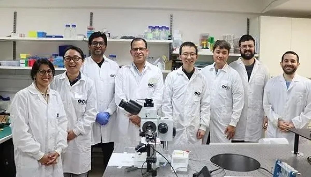
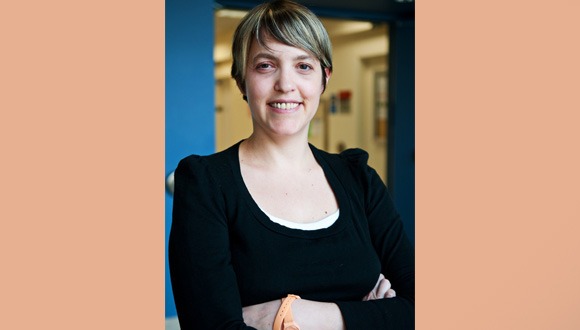
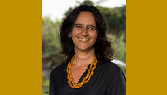
The researchers (from left to right): Dr. Maya Geva-Sagiv, Prof. Yuval Nir and Prof. Itzhak Fried

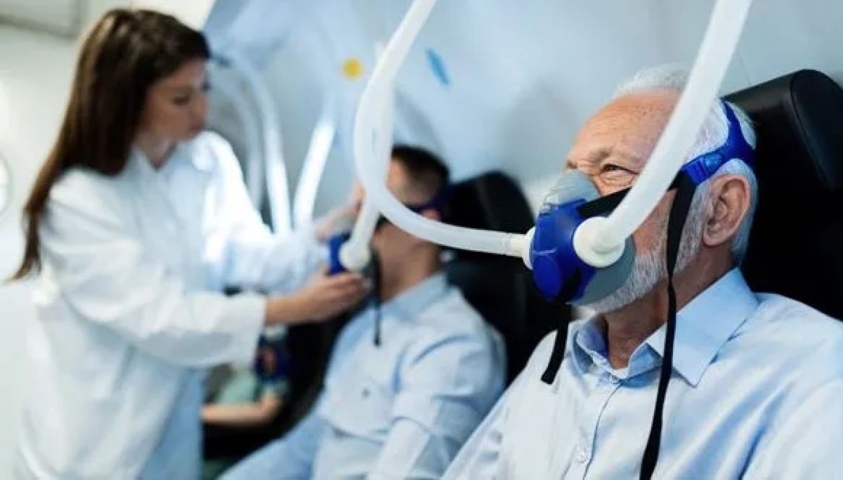
“This study was made possible by a rare group of 18 patients with epilepsy at the UCLA Medical Center,” says Prof. Nir. “Prof. Fried implanted electrodes in these patients’ brains to try and pinpoint the areas that cause their epileptic seizures, and they volunteered to take part in a study investigating the effects of deep-brain stimulation during sleep. Close work with expert neurologists led by Prof. Dawn Eliashiv at UCLA enabled our team to integrate advanced brain stimulation in the research. Thus, we were able to test, for the first time in humans, the long-held hypothesis – that coordinated activity of the hippocampus and cerebral cortex during sleep is a critical mechanism in consolidating memories.”

“Moreover, we improved memory consolidation through a special stimulation protocol that enhanced synchronization between these two areas in the brain. Intervention during sleep represents a unique approach that can be further developed in the future to provide hope for people with memory impairments such as dementia.”


“In this study we directly examined the role of neural activity and electrical brain waves during sleep. Our goal was to enhance the natural mechanisms at play, to discover exactly how sleep assists in stabilizing memories.” – Dr. Maya Geva-Sagiv


Unraveling Mechanism

“We know that a good night’s sleep is critical for the consolidation of long-lasting memories, but so far, we had little evidence regarding the precise processes that are at work during human sleep,” explains Dr. Maya Geva-Sagiv. “In this study we directly examined the role of neural activity and electrical brain waves during sleep. Our goal was to enhance the natural mechanisms at play, to discover exactly how sleep assists in stabilizing memories.”

The researchers developed a deep-brain stimulation system that improves electrical communication between the hippocampus – a deep-brain region involved in acquiring new memories, and the frontal cortex – where memories are stored for the long term. By monitoring activity in the hippocampus during sleep, the system enables precisely timed delivery of electrical stimulation to the frontal cortex.

The study’s participants completed two memory tests, and their performance was compared after two different nights – one undisturbed and one with deep-brain stimulation. On both occasions, they were asked in the morning to recognize famous persons whose pictures they had been shown the previous evening. The study found that deep-brain stimulation significantly improved the accuracy of their memory.


“To our surprise, we also discovered that the intervention did not significantly increase the number of right answers given by participants, but rather reduced the number of wrong answers. This suggests that sleep sharpens the accuracy of our memory…”   – Prof. Yuval Nir


Sharpening Memory Accuracy

“We found that our method had a beneficial effect on both brain activity during sleep and memory performance,” says Prof. Fried. “All patients who had received synchronized stimuli to the frontal cortex demonstrated better memory performance, compared to nights of undisturbed sleep. The control group, which received similar yet unsynchronized stimuli, showed no memory improvement. Our deep-brain stimulation method is unique because it is close-looped – stimuli are precisely synchronized with hippocampal activity. In addition, we monitored the stimuli’s impact on brain activity at a resolution of individual neurons.”

“Our findings support the hypothesis that precise coordination between sleep waves assists communication between the hippocampus that takes in new memories, and the frontal cortex that stores them for the long term,” adds Prof. Nir.

“To our surprise, we also discovered that the intervention did not significantly increase the number of right answers given by participants, but rather reduced the number of wrong answers. This suggests that sleep sharpens the accuracy of our memory, or in other words, it removes various distractions from the relevant memory trace.”   

  The study was supported by grants from the US National Institutes of Health (NIH), the European Research Council (ERC), the US National Science Foundation (NSF), the US-Israel Bilateral Science Foundation (BSF), and the Human Frontier Science Program (HFSP). The paper’s other co-authors are: Prof. Dawn Eliashiv, Dr. Emily Mankin, Natalie Cherry, Guldamla Kalender, and Dr. Natalia Tchemondanov of UCLA, and Dr. Shdema Epstein from Tel Aviv University.

Prof. Yosef Shiloh Elected as International Member of US National Academy of Sciences

Renowned Tel Aviv University Professor Emeritus recognized for pioneering cancer research and advocacy for rare genetic disorders.

Professor Emeritus Yosef Shiloh from TAU’s Sackler Faculty of Medicine was elected an international member of the US National Academy of Sciences (NAS). The Academy includes approximately 2,500 American scientists from all fields of science and another approximately 500 foreign scientists from all over the world. Prof. Shiloh is the 43rd Israeli researcher elected to NAS, alongside Nobel laureates Prof. Ada Yonath, Prof. Dan Shechtman and Prof. Aaron Ciechanover. Prof. Shiloh is the Incumbent of the David and Inez Myers Chair for Cancer Genetics in the Department of Human Molecular Genetics and Biochemistry in the Sackler Faculty of Medicine at Tel Aviv University.


Prof. Shiloh received the exciting news while attending a conference in Boston. He has been the recipient of the EMET Prize, the Israel Prize, and the Clowes Award for Outstanding Cancer Research – the most important prize awarded by the American Association for Cancer Research, which has thousands of cancer researchers as members. Being elected to the Academy is a particular honor, as it reflects widespread recognition by all the members of the Academy, from a variety of scientific fields. The acceptance bar for non-American members is particularly high, adding to the prestige of “international” members.


The US National Academy of Sciences advises the American government and nation on matters of science, engineering and medicine, based on a charter granted to it by Congress and signed by President Abraham Lincoln in 1863. Membership in the Academy is lifelong, and up to 120 scientists from the USA and up to 30 foreign scientists are elected to the Academy each year.


Prof. Shiloh: “It is a great honor and I thank the Academy members for recognizing our work. NAS is a body whose opinion is heard and given consideration and I hope that the opinion of the Israeli Academy of Sciences and Humanities, of which I am a member, will be heard here in a similar way. In the US, the president, the administration and the public listen to what the Academy says, and hence the weight that the Americans attribute to membership in this institution.”


Prof. Karen Avraham, Dean of the Sackler Faculty of Medicine:” This is a tremendous honor for us in the Department and the Faculty of Medicine at Tel Aviv University. Prof Yosef Shiloh’s research has made a seminal and remarkable contribution in the area of a rare but devastating genetic disease, ataxia-telangiectasia (A-T), with far-reaching implications for DNA repair and cancer. Most compelling, what drove Prof. Shiloh every step of the way was his compassion for the patients. The main theme of his work can be summarized in the title of a lecture that he has delivered to numerous audiences over the years: “Investigation of Rare Genetic Disorders: A Mission for Human Welfare and a Steppingstone in Understanding our Biology”. Prof. Shiloh continues to explore the connection between A-T, neurodegeneration and aging in search of new treatment modalities for A-T, as well as to devote his efforts to educating the public about the medical and social implications of the genome revolution.”


Next year Prof. Shiloh will participate in a ceremony in honor of the members selected this year, which will be held at the Academy House in Washington, DC.

Tel Aviv University Researchers Present New Treatment for Ovarian Cancer

Using RNA-based nanodrugs the researchers achieve 80% survival rate in lab models.

Ovarian cancer ranks fifth in cancer deaths among women, accounting for more deaths than any other cancer of the female reproductive system. In a study conducted at Tel Aviv University researchers used protein CKAP5 (cytoskeleton-associated protein) for the first time as a therapeutic target for RNA-based nanodrugs. After identifying a genetically unstable mutation resistant to both chemotherapy and immunotherapy in the tissues of ovarian cancer, the researchers targeted these cells with lipid nanoparticles containing RNA for silencing CKAP5 – causing the cells to collapse and achieving an 80% survival rate in animal models.


“The lipid nanoparticles developed by Prof. Peer enabled us for the first time to silence [the CKAP5] protein through targeted delivery of an RNA drug. We proved that CKAP5, a protein responsible for the cell’s stability, can be silenced, and that this procedure collapses and destroys the entire cancer cell.” – Dr. Sushmita Chatterjee


Targeted Delivery of RNA Drug

The breakthrough was achieved by a TAU research team led by Prof. Dan Peer of The Shmunis School of Biomedicine and Cancer Research, a global pioneer in the development of RNA-based drugs, Head of the Laboratory of Precision Nanomedicine, and TAU’s VP for R&D; and by Dr. Sushmita Chatterjee, post-doctoral student from India at Prof. Peer’s lab, in collaboration with Prof. David Sprinzak of The George S. Wise Faculty of Life Sciences and Prof. Ronen Zaidel-Bar of the Sackler Faculty of Medicine. The study was funded by the Rivkin Foundation for Ovarian Cancer Research and the Shmunis Family Foundation. The results were published in the leading scientific journal Science Advances.

“The protein CKAP5 has never been studied with relation to the fight against cancer, simply because there was no known way to silence it,” explains Dr. Chatterjee. “The lipid nanoparticles developed by Prof. Peer enabled us for the first time to silence this protein through targeted delivery of an RNA drug. We proved that CKAP5, a protein responsible for the cell’s stability, can be silenced, and that this procedure collapses and destroys the entire cancer cell.”


Prof. Dan Peer

“Something Like a Dominoes Game”

At the second stage of the study the researchers tested the new CKAP5-silencing RNA drug on 20 types of cancer. Some cancer cells proved more sensitive than others to this procedure. Cancers displaying high genetic instability, which are usually highly resistant to chemotherapy, were found to be especially sensitive to the silencing of CKAP5.


“As researchers, we are involved in something like a dominoes game: we always look for the one piece in the cancer’s structure that is so important, that if we pull it out the entire cell will collapse. CKAP5 is such a domino piece, and we are already working on more applications (…)” – Prof. Dan Peer


“All cancer cells are genetically unstable,” says Dr. Chatterjee. “Otherwise, they would be healthy, not cancerous. However, there are different levels of genetic instability. We found that cancer cells that are more unstable, are also more affected by damage to CKAP5.  Our drug pushed them to their limit, and essentially destroyed their structure. Our idea was to turn the trait of genetic instability into a threat for these cells, by using RNA to silence the flawed protein. We demonstrated for the first time that CKAP5 can be used to kill cancer cells, and then observed the biological mechanism that causes the cancer cells to collapse in the protein’s absence.”

Equipped with these insights, the researchers tested the new drug in an animal model for ovarian cancer, achieving a survival rate of 80%.

“We chose ovarian cancer because it’s a good target,” explains Prof. Peer. “While highly resistant to both chemotherapy and immunotherapy, this type of cancer is very sensitive to the silencing of CKAP5. It should be emphasized that the CKAP5 protein is a new target in the fight against cancer. Targeting cell division is not new, but using RNA to target proteins that make up the cell’s skeleton (cytoskeleton) – this is a new approach and a new target that must be further investigated. As researchers, we are involved in something like a dominoes game: we always look for the one piece in the cancer’s structure that is so important, that if we pull it out the entire cell will collapse. CKAP5 is such a domino piece, and we are already working on more applications, this time in blood cancers.”

“Family Smoking” on The Porch

Six out of ten children whose parents restrict their smoking to the porch are at risk for being harmed by tobacco smoke.

Many parents think that they are protecting their children by smoking on the porch or next to the window in a room. However, a new study by Tel Aviv University finds that, in contrast to such beliefs, restricting smoking to the porch does not protect most children from exposure to tobacco smoke. The research team tested for the presence of nicotine in the hair of children whose parents restrict their smoking to the porch or outside the house. Their findings are worrisome: nicotine was found in the hair of six out of ten children.

The researchers emphasize that “in Israel, home porches should be regarded as part of the environment of the home. Smoking next to a window or in another specific place in the home does not protect most children from exposure. Our recommendations are unequivocal: to reduce children’s exposure to tobacco smoke, smoking should be entirely avoided within a range of ten meters from the house. Likewise, in open areas, smokers should maintain a distance of at least ten meters from the children.”


“The Israeli situation is of great concern because in many cases, porches in Israel are directly adjacent to the living areas and may even be partially open some of the time (…) The parents mistakenly believe that the porch offers a ‘safe’ place to smoke.” Prof. Leah (Laura) Rosen


The Porch is No ‘Safe’ Place

The study was led by Prof. Leah (Laura) Rosen from the School of Public Health in Sackler Faculty of Medicine, Tel Aviv University. Also participating in the study: Prof. David Zucker from the Department of Statistics and Data Science, Hebrew University, Jerusalem; Dr. Shannon Gravely from the Department of Psychology, Waterloo University, Canada; Dr. Michal Bitan from the Computer Science Department, the College of Management; Dr. Ana Rule from the Department of Health and Environmental Engineering, Johns Hopkins Bloomberg School of Public Health, Baltimore; and Dr. Vicki Meyers from the Gertner Institute for Epidemiology and Public Policy Research, Sheba Medical Center. The study was published in the International Journal of Environmental Research and Public Health.

In the first stage of the study (published about two years ago), the research team studied hair samples of the children of smoking parents for the presence of nicotine. This provides an estimate of their exposure to tobacco smoke over the past months. It was found that 70% of the children of smoking parents had measurable hair nicotine.

In the current stage of the study, the researchers examined the data by the location of parental smoking. Analysis of the data showed that in families in which the parents restricted their smoking to the porch or outdoors, 62% of the children were still exposed to tobacco smoke.

Prof. Leah (Laura) Rosen

“It is known that smoking outside the house, even when the doors and windows are fully closed, does not completely protect children from exposure to tobacco smoke,” says Prof. Rosen. “The Israeli situation is of great concern because in many cases, porches in Israel are directly adjacent to the living areas and may even be partially open some of the time. The proximity allows smoke to drift from those areas to the interior of the house. The parents mistakenly believe that the porch offers a ‘safe’ place to smoke.”

“In fact, the children are likely to be directly exposed when they come out to the porch and someone is smoking, or when smoke drifts into the house. Once in the home, the smoke is absorbed into the environment, for example, into the furniture or walls or rugs, and is then gradually discharged into the air over weeks or months.”

“Further, this residual smoke, known as third hand smoke, can be absorbed into the body from the environment via swallowing or through the skin, especially among infants and small children. In addition, smoking parents transmit the toxins from the tobacco smoke on their skin, on their hands, in their hair, on their clothing. Therefore, it is recommended to brush teeth, wash hands, and change clothes after smoking, before contact with children.”


“85% of tobacco smoke is invisible, and our sense of smell is not reliable, so many parents mistakenly believe that they are protecting their children, while in fact they are exposing them to substantial health risks.” Prof. Leah (Laura) Rosen


Plea to Israel’s Health Ministry

Prof. Rosen notes that this new information is directly relevant to Case 1416/21 on neighbor smoking, currently being heard in the Supreme Court. The appeal against the Ministries of the Environment, Health, and Interior concerns the tobacco smoke that penetrates apartments as an environmental hazard, a claim that is supported by the definition of an environmental hazard in the Clean Air Law, the Hazard Prevention Law, and the Penal Code.

Prof. Rosen: “The results of this study show that among smoking families, restricting smoking to the porch does not protect most children from exposure to tobacco smoke. Therefore, the Health Ministry’s approach, which opposes protection for individuals from smoke incursion into their own homes to protect the smokers’ children, does not protect the children of smokers, and in addition it can cause substantial harm to neighbors and the children of neighbors.  We ask the Health Ministry to reconsider its stand in light of these findings.”

“The State of Israel must make the reduction of parental smoking a national goal and invest the appropriate resources in this issue. Unfortunately, there are many misconceptions regarding when and how the exposure occurs. 85% of tobacco smoke is invisible, and our sense of smell is not reliable, so many parents mistakenly believe that they are protecting their children, while in fact they are exposing them to substantial health risks. As a society, we must safeguard citizens and distance everyone from the risks of tobacco smoke exposure, especially infants and children, pregnant women, and all vulnerable populations,” concludes Prof. Rosen.

Tiny Robot Navigates in Physiological Environment and Captures Targeted Damaged Cells

Meet the hybrid micro-robot: innovative technology only 10 microns across.

Researchers at Tel Aviv University have developed a hybrid micro-robot, the size of a single biological cell (about 10 microns across), that can be controlled and navigated using two different mechanisms – electric and magnetic. The micro-robot is able to navigate between different cells in a biological sample, distinguish between different types of cells, identify whether they are healthy or dying, and then transport the desired cell for further study, such as genetic analysis. The micro-robot can also transfect a drug and/or gene into the captured targeted single cell. According to the researchers, the development may help promote research in the important field of ‘single cell analysis’, as well as find use in medical diagnosis, drug transport and screening, surgery, and environmental protection.

Inspired by Biological Micro-swimmers

The innovative technology was developed by Prof. Gilad Yossifon from the School of Mechanical Engineering and Department of Biomedical Engineering at Tel Aviv University and his team: post-doctoral researcher Dr. Yue Wu and student Sivan Yakov, in collaboration with Dr. Afu Fu, Post-doctoral researcher, from the Technion, Israel Institute of Technology. The research was published in the journal Advanced Science.


“Developing the micro-robot’s ability to move autonomously was inspired by biological micro-swimmers, such as bacteria and sperm cells. This is an innovative area of research that is developing rapidly, with a wide variety of uses in fields such as medicine and the environment, as well as a research tool.” – Prof. Gilad Yossifon


Prof. Gilad Yossifon explains that micro-robots (sometimes called micro-motors or active particles) are tiny synthetic particles the size of a biological cell, which can move from place to place and perform various actions (for example: collection of synthetic or biological cargo) autonomously or through external control by an operator. According to Prof. Yossifon, “developing the micro-robot’s ability to move autonomously was inspired by biological micro-swimmers, such as bacteria and sperm cells. This is an innovative area of research that is developing rapidly, with a wide variety of uses in fields such as medicine and the environment, as well as a research tool”.


WATCH: The Hybrid Micro-Robot


As a demonstration of the capabilities of the micro-robot the researchers used it to capture single blood and cancer cells and a single bacterium, and showed that it is able to distinguish between cells with different levels of viability, such as a healthy cell, a cell damaged by a drug, or a cell that is dying or dying in a natural ‘suicide’ process (such a distinction may be significant, for example, when developing anti-cancer drugs).

After identifying the desired cell, the micro-robot captured it and moved the cell to where it could be further analyzed. Another important innovation is the ability of the micro-robot to identify target cells that are not labeled – the micro-robot identifies the type of cell and its condition (such as degree of health) using a built-in sensing mechanism based on the cell’s unique electrical properties.

Effective in Physiological Environments

“Our new development significantly advances the technology in two main aspects: hybrid propulsion and navigation by two different mechanisms – electric and magnetic,” explains Prof. Yossifon. “In addition, the micro-robot has an improved ability to identify and capture a single cell, without the need for tagging, for local testing or retrieval and transport to an external instrument. This research was carried out on biological samples in the laboratory for in-vitro assays, but the intention is to develop in the future micro-robots that will also work inside the body – for example, as effective drug carriers that can be precisely guided to the target”.


“… the technology will support the following areas: medical diagnosis at the single cell level, introducing drugs or genes into cells, genetic editing, carrying drugs to their destination inside the body, cleaning the environment from polluting particles, drug development, and creating a ‘laboratory on a particle’ – a microscopic laboratory designed to carry out diagnostics in places accessible only to micro-particles.” – Prof. Gilad Yossifon


The researchers explain that the hybrid propulsion mechanism of the micro-robot is of particular importance in physiological environments, such as found in liquid biopsies: “The micro-robots that have operated until now based on an electrical guiding mechanism were not effective in certain environments characterized by relatively high electrical conductivity, such as a physiological environment, where the electric drive is less effective. This is where the complementary magnetic mechanism come into play, which is very effective regardless of the electrical conductivity of the environment”.

Prof. Yossifon concludes: “In our research we developed an innovative micro-robot with important capabilities that significantly contribute to the field: hybrid propulsion and navigation through a combination of electric and magnetic fields, as well as the ability to identify, capture, and transport a single cell from place to place in a physiological environment. These capabilities are relevant for a wide variety of applications as well as for research. Among other things, the technology will support the following areas: medical diagnosis at the single cell level, introducing drugs or genes into cells, genetic editing, carrying drugs to their destination inside the body, cleaning the environment from polluting particles, drug development, and creating a ‘laboratory on a particle’ – a microscopic laboratory designed to carry out diagnostics in places accessible only to micro-particles.”

Hyperbaric Treatment More Effective than Medicines for Fibromyalgia Caused by Head Injury

Researchers say “results were dramatic” for patients who underwent hyperbaric oxygen therapy.

Researchers from Tel Aviv University compared treatment with a dedicated protocol of hyperbaric oxygen therapy (HBOT) to the pharmacology (drugs) treatment available today for patients suffering from fibromyalgia, a chronic pain syndrome, caused by traumatic brain injury (TBI). Their findings showed that dedicated hyperbaric oxygen therapy is much more effective in reducing pain than the drug treatment and ended up healing two out of five of the participants in the study.

Chronic Pain Syndrome

The study was conducted by researchers from Tel Aviv University’s Sackler Faculty of Medicine, led by Prof. Shai Efrati, MD, from the Sagol Center for Hyperbaric Medicine and Research at the Shamir Medical Center, and Prof. Jacob Ablin, MD, from the Tel Aviv Sourasky Medical Center. The results of the study were published in the journal PLOS One.  


“At the end of the treatment, two out of five patients in the hyperbaric treatment group showed such a significant improvement that they no longer met the criteria for fibromyalgia. In the drug treatment group, this did not happen to any patient.” Prof. Shai Efrati


“Fibromyalgia is a chronic pain syndrome, from which between 2% – 8% of the population suffers,” explains Prof. Shai Efrati. “Until 15-20 years ago, there were doctors who believed that it was a psychosomatic illness and recommended that patients with chronic pain seek mental health care. Today we know that it is a biological illness, which damages the brain’s processing of the signals received from the body. When this processing is malfunctioning, you feel pain without any real damage in related locations.”

“Fibromyalgia can be induced by variable triggers – from certain infections, as we have recently seen in post-COVID patients, through post-traumatic stress syndrome to head injuries. We wanted to test whether the new protocols of hyperbaric medicine can provide better results than pharmacological medicine, for patients in whom the fibromyalgia was induced by traumatic brain injury.”


Prof. Shai Efrati


Dramatic Results

Hyperbaric medicine is a form of treatment in which the patients stay in special chambers where the pressure is higher than the atmospheric pressure at sea level, and where the patients breathe 100% oxygen. Hyperbaric medicine is considered safe, used in many places including Israel, and is already used to treat a long list of medical conditions.

In recent years, scientific evidence has been accumulating that certain, newly developed, dedicated hyperbaric treatment protocols can lead to the growth of new blood vessels and neurons in the brain.


“Overall, existing treatments are not good enough. [Fibromyalgia] is a chronic disease that significantly affects the quality of life, including young people, and hyperbaric medicine meets an acute need of these patients.” Prof. Jacob Ablin


In their current study, the researchers from Tel Aviv University recruited 64 Israelis aged 18 and older who suffered from fibromyalgia as a result of a head injury, and randomly divided them into two groups: one group was exposed to 100% pure oxygen at a pressure of two atmospheres for 90 minutes (with fluctuations in oxygen during the treatment every 20 minutes), five days a week, for three months. The second group received the conventional pharmacological treatment (i.e., the drugs pregabalin, which is known under the trade name “Lyrica”, and duloxetine, which is better known as “Cymbalta”).

“The results were dramatic,” says Prof. Efrati. “At the end of the treatment, two out of five patients in the hyperbaric treatment group showed such a significant improvement that they no longer met the criteria for fibromyalgia. In the drug treatment group, this did not happen to any patient. Furthermore, the average improvement in the pain threshold tests was 12 times better in the hyperbaric group compared to the medication group. And in terms of quality-of-life indicators, as reported by the patients, we saw significant improvements in all the indicators among the patients who received hyperbaric treatment.”

Meets Acute Need

“Today’s accepted treatment for fibromyalgia includes pharmacologic and non-pharmacologic components,” says Prof. Ablin. “with respect to the pharmacologic approach, these drugs are not very effective and therefore the emphasis is on the non-pharmacological side, that is, on external correction of pain processing within the nervous system. Currently used recommendations includes aerobic activity, hydrotherapy, cognitive-behavioral therapy and movement-based therapies such as Tai Chi. In addition, quite a few patients request treatment with medical cannabis, and for some it helps.”


“In the group that received hyperbaric treatment, you could see the repair of the brain tissue, while in the control group there was only an attempt to relieve the pain – without treating the damaged tissue – and of course the medication group experienced the side effects associated with drug treatment.” Prof. Shai Efrati


“Overall, existing treatments are not good enough. [Fibromyalgia] is a chronic disease that significantly affects the quality of life, including young people, and hyperbaric medicine meets an acute need of these patients. Of course, these are preliminary studies, and we must follow and see what effect the medical protocol has on the patients after one, two and three years – and if it is necessary to maintain the positive results with further exposure to hyperbaric sessions.”

Looking to Cure

According to Prof. Efrati, the importance of the research is in healing the damaged brain tissue – and not in treating its superficial symptoms: “In the group that received hyperbaric treatment, you could see the repair of the brain tissue, while in the control group there was only an attempt to relieve the pain – without treating the damaged tissue – and of course the medication group experienced the side effects associated with drug treatment. This is a difference in approach: to cure instead of just treating the symptoms.”

“We assessed the improvement of the participants in the hyperbaric group more than a week after the last hyperbaric session. More follow-up studies are needed to see the duration of the beneficial effect of the treatment and if and for whom additional treatment will be needed. Our goal as doctors is not only to treat the symptoms but, to the extent possible, also to treat the source of the problem, thus improving the quality of life of fibromyalgia patients.”

“It is important to emphasize that the dedicated hyperbaric oxygen treatment protocol found to be effective is only available in medical centers that have licensed hyperbaric chambers. Be careful of so-called ‘private chambers’, since these cannot provide the therapeutic protocol found to be effective, and they are not regulated or approved for medical use,” cautions Prof. Efrati.

Promoting Women in Medicine

New enrichment program created for and by Tel Aviv University’s female medical students.

Women in Medicine (WIM) is a new program created for and by TAU’s female medical students. The semester-long program kicks off on March 15 and aims to offer open dialogue and a community of female students from the Sackler Faculty of Medicine where they can receive advice, mentorship and support. 

The participants come from different study programs and years. Together, they will be listening to lectures by female faculty members (all successful women in medicine and most of them renowned doctors in various fields) who will share their own points of view on the unique challenges faced by women in the profession, accompanied by a wealth of knowledge acquired through their own personal experience. Outside experts will contribute as well, and topics to be covered include clinical research, entrepreneurship, and technology. The group will meet with a hospital director; attend a public speaking workshop where they will learn how to crush it in job interviews; participate in a LinkedIn workshop at LinkedIn’s offices and much more. The in-person aspect of the meetings is key to create open dialogue and to facilitate meaningful connections between the women.

Orr Erlich, one of the co-founders of WIM, filled us in with more details. 

Orr, why do we need a separate program to promote women in medicine?

“The majority of TAU’s medical students today are female. Sadly, this encouraging number is not reflected in women’s reprensentation in many healthcare management positions in Israel. We want to set our female students up for success, and we will do so by organizing a full semester of bi-weekly meetings, whereby each meeting is divided into two parts: the first part will be instructive and go in-depth on issues that are important to our conduct as women in this very demanding profession, both on a personal and professional level. Established professionals will share from their knowledge and lessons learnt on topics like entrepreneurship; integration into research and academic advancement; management skills and teamwork; self-presentation; public speaking; resilience and dealing with difficult parts of the profession; cultivating negotiation skills; achieving a work-home balance and the importance of networking. The students will also be made aware of advantages they may have as women. The second part of the meetings will be practical workshops on the topics discussed earlier.”

How did you select the participants for the pilot group?

“All the participants are female medical students from our University, and they come from a variety of programs (six-year and four-year, female medical students doing a PhD or MPH) and from all degree-stages. It is very important for us to maintain this diversity and to bring female students from different backgrounds, stages of life and interests (for example research or innovation) and with different strengths and abilities.”

What will the participants take away from this program?

“WIM will enable each participant to seek out mentorship and share professional concerns and aspirations with likeminded women. We want the women to emerge from the program with raised self-esteem, with the understanding that they can realize their dreams and with the knowledge that they are in charge of their own medical careers.”

Stay updated on application dates for new program cycles and events that will be open for the public and oganized throughout the year on Women in Medicine’s social media channels (Instagram, Facebook, LinkedIn). 

Learn more about WIM

Would you like to get in touch with the organizers, or perhaps you’d be interesting in coming to speak to the group?

Send an email to: [email protected] 

#TAU_WOMEN_POWER

Meet female researchers from Tel Aviv University who received the prestigious European Research Council (ERC) grants this year.

Four female researchers from Tel Aviv University received the prestigious European Research Council (ERC) grants, aimed to help promising mid-career researchers achieve their research goals. We spoke with the winners to hear how they feel about the award and hear any advice they may have for other aspiring researchers.

Prof. Miri Yemini | The School of Education at TAU’s Faculty of Humanities 

In her research, Yemini explores how young people from different backgrounds in different countries understand and appropriate global citizenship.

How did you feel when you heard that you’d be awarded the grant?

“I was very excited when I received the news about the grant. At the same time, I understand that winning the grant is just the beginning. It marks a long road ahead, with research and action, fueled by curiosity and perseverance.”

This year, the majority of TAU’s ERC grant recipients are female. How do you feel about that?

“I am happy for all the recipients and look forward to future updates on their research and results. I am eager to see more women in senior positions at Israeli universities, both in academic and administrational positions. I would also like to see a more diverse academia in terms of gender, religion, ethnicity, first-generation academics and more. We still have a long way to go in this respect.”

Do you have a personal message for young female researchers at the beginning of their career?

“To win a grant, you first need to submit. For me, it has been helpful to filter out fears and hesitations and focus on what interests and excites me research-wise. It is also important to bear in mind that behind most such achievements are at least twice as many failures – and these don’t tend to make the headlines.”


Prof. Miri Yemini

Prof. Limor Landsman | The Cell and Developmental Biology Department of TAU’s Sackler Faculty of Medicine

Landsman researches the function of beta cells, cells that regulate insulin production, crucial for blood sugar control and for the prevention of diabetes. Her team studies how beta-cell function and mass are established and maintained in healthy individuals and why they are lost in instances of diabetes.

How did you feel when you heard that you won the grant?

“I felt happy and proud, but most of all relieved that my lab will be funded in the coming years, so that I can continue to advance our research in a direction that I believe will lead to new insights into diabetes.”

This year, most TAU’s ERC grant recipients are female. How do you feel about that?

“I am proud to be part of an academic institution that advocates excellence, of both women and men.”
 

Do you have a personal message for young female researchers at the beginning of their career?

“What I like about research is that nothing is known in advance, for better and for worse. That’s why I think it’s important not to be afraid to try, even if the chances are low. Did it work? Great! If it didn’t – allow yourself to be upset for a day or two and then dust if off and try a different direction.” 


Prof. Limor Landsman


Prof. Hila Shamir | Tel Aviv University – The Buchmann Faculty of Law

Shamir teaches and researches Employment, Labor, Immigration, and Welfare Law with a focus on issues of human trafficking, gender equality, informal work, and the law of global value chains. She is a second time ERC grant winner, and as part of her first ERC grant, she established the research group TraffLab: Labor Perspective to Human Trafficking.

In her current research, Shamir examines efforts to promote workers’ rights in global supply and production chains.

How did you feel when you heard that you’d be awarded the grant?

“I was very excited. This is the second time I receive an ERC grant, and I assumed that my chances were low as a result. So, the surprise of receiving the grant this year was great. I also feel an accompanying weight of responsibility and the desire to ensure efficient use of such a significant amount.”

This year, most of TAU’s ERC grant recipients are female. How do you feel about that?

“Very happy and not at all surprised. There are wonderful female researchers in the Israeli academia, and it is great to see their work receiving international recognition.”

Do you have a personal message for young female researchers at the beginning of their career?

“Don’t be afraid to submit a grant application, even if it may seem intimidating initially. Do invest some time on the submission, as a grant of this scale has the potential to greatly change the way we conduct research: it makes it possible to build a large research group; secure funding for researchers at the beginning of their career and enables significant research scope with a correspondingly strong potential to influence. Even if you should not end up winning, the attempt to think big and imagine research on such a scale is valuable in and by itself and could bear fruits further down the line. Good luck!”


Prof. Hila Shamir

We wish our female rockstars congrats and good luck with their current research!


 Established in 2007 and annually awarded by the European Union, the ERC research grant is considered one of the most prestigious and important ones in the world of science. The four main types of ERC grants are: ERC Starting Grant, ERC Consolidator Grant, ERC Advanced Grant and the ERC Synergy Grant.

Researchers Discover Mechanism that Facilitates Formation of Brain Metastases

Findings could help predict metastatic recurrence in the brain and a worse prognosis.

Brain metastases are one of the deadliest forms of cancer metastasis, with grave survival rates of less than one year in many cases. The incidence of brain metastases has been increasing in recent years and developing better therapeutic strategies for brain metastasis is an urgent need. In a new study from Tel Aviv University, researchers identified and characterized a new mechanism that facilitates the formation of brain metastases and found that impairing this mechanism significantly reduced the development of brain metastases in lab models.


“The findings establish LCN2 as a new prognostic marker and a potential therapeutic target.” Prof. Neta Erez


On the Radar: LCN2

The research was led by Prof. Neta Erez, head of the laboratory for the biology of tumors from the Department of Pathology at the Sackler Faculty of Medicine, and members of her team: Omer Adler, Yael Zeit, and Noam Cohen, in collaboration with Prof. Shlomit Yust Katz from Rabin Medical Center (Beilinson Hospital) and Prof. Tobias Pukrop from Regensburg Hospital, Germany. The study was supported by the Melanoma Research Alliance (MRA), the Cancer Biology Research Center at Tel Aviv University, the Personalized Medicine Program of the Israel Science Foundation (ISF IPMP) and the German Cancer Research Foundation (DFG), and was published in the journal Nature Cancer.

In this new study, the researchers show that Lipocalin-2 (LCN2) [a protein which in humans is encoded by the LCN2 gene] is a key factor in inducing neuroinflammation in the brain. Moreover, the researchers found that high LCN2 levels in patients’ blood and brain metastases from several types of cancer are associated with disease progression and reduced survival.

LCN2 is a secreted protein that functions in the innate immune system and was originally discovered due to its ability to bind iron molecules and as part of the inflammatory process in fighting bacterial infection. LCN2 is produced by a large variety of cells and was shown to be involved in multiple cancer-related processes.

“Our findings reveal a previously unknown mechanism, mediated by LCN2, which reveals a central role for the mutual interactions between immune cells recruited to the brain (granulocytes) and brain glial cells (astrocytes) in promoting inflammation and in the formation of brain metastases. The findings establish LCN2 as a new prognostic marker and a potential therapeutic target,” says Prof. Neta Erez.


“In blood and tissue samples from patients with brain metastases from three types of cancer, blood LCN2 levels were correlated with disease progression and with shorter survival, which positions LCN2 as a potential prognostic marker for brain metastases.” Prof. Neta Erez


LCN2 as a Predictive Marker for Brain Metastases

The researchers used models of melanoma and breast cancer brain metastases to reveal the mechanism by which neuroinflammation is activated in the metastatic niche in the brain.

“We show that signals secreted into the blood from the primary tumor stimulate pro-inflammatory activation of astrocytes in the brain. The astrocytes promote the recruitment of inflammatory cells from the bone marrow (granulocytes) into the brain, and they in turn become a main source of signaling by LCN2,” explains Prof. Erez.

“We demonstrated the importance of LCN2 for the development of metastases by genetically inhibiting its expression in mice, which resulted in a significant decrease in neuroinflammation and reduced brain metastases. Moreover, in blood and tissue samples from patients with brain metastases from three types of cancer, blood LCN2 levels were correlated with disease progression and with shorter survival, which positions LCN2 as a potential prognostic marker for brain metastases.”

Prof. Erez adds: “We analyzed the LCN2 protein levels in the blood and cerebrospinal fluid (CSF) of mice with brain metastases and found that LCN2 levels increased greatly in mice with melanoma and breast cancer metastases compared to healthy mice. Importantly – an increase in blood LCN2 preceded the detection of brain metastases by MRI. Furthermore, the mice in which LCN2 levels were very high developed brain metastases later, further establishing LCN2 as a predictive marker for brain metastases.”

The researchers also examined whether LCN2 is elevated in the blood of melanoma patients at the time of initial diagnosis, and whether it can be a prognostic factor. The findings indicated that patients with melanoma had significantly higher levels of LCN2 in their blood compared to samples from healthy individuals. Strikingly, patients who developed brain metastases displayed significantly higher levels of LCN2 even before the diagnosis of the metastases, and high levels of LCN2 in the blood correlated with worse survival.

“We have identified a new mechanism in which LCN2 mediates the communication between immune cells from the bone marrow and supporting cells in the brain, activates inflammatory mechanisms and thus helps the progression of metastatic disease in the brain, and demonstrated its importance. The functional and prognostic aspects of LCN2 that we have identified in brain metastases in mouse models as well as in cancer patients suggest that targeting LCN2 may be an effective therapeutic strategy to delay or prevent the recurrence of brain metastases,” summarizes Prof. Erez.

Light Pollution is Killing Desert Rodents

New study shows that artificial light at night can be harmful to ecosystems, biodiversity, and human health.

A new study from Tel Aviv University’s School of Zoology tested the impact of prolonged low-intensity light pollution on two species of desert rodents: the diurnal golden spiny mouse, and the nocturnal common spiny mouse. The findings were highly disturbing: on two different occasions, entire colonies exposed to ALAN (Artificial Light At Night) died within days, and reproduction also decreased significantly compared to control groups. According to the researchers, the results show clearly for the first time that light pollution can be extremely harmful to these species, and suggest they may be harmful to ecosystems, biodiversity, and even human health.


“According to latest studies, about 80% of the world’s human population is exposed to ALAN, and the area affected by light pollution grows annually by 2-6%. In a small and overcrowded state like Israel, very few places remain free of light pollution.” Hagar Vardi-Naim


Humans Changed the Rules

The study was led by Prof. Noga Kronfeld-Schor, Chief Scientist of Israel’s Ministry of Environmental Protection, and PhD student Hagar Vardi-Naim, both from TAU’s School of Zoology and the Steinhardt Museum of Natural History.  The paper was published in Scientific Reports.

“We have been studying these closely related rodent species for years.  They both live in Israel’s rocky deserts: the golden spiny mouse (Acomys russatus) is diurnal [active during the day], and the common spiny mouse (A. cahirinus) in nocturnal [active during the night],” explains Prof. Kronfeld-Schor. “The two species share the same natural habitat but use it at different times to avoid competition. By comparing closely related species that differ in activity times, we gain new insights into the biological clock and its importance to the health of both animals and humans.”

Hagar Vardi-Naim notes that, “in most species studied to date, including humans, the biological clock is synchronized by light. This mechanism evolved over millions of years in response to the daily and annual cycles of sunlight – day and night and their varying lengths that correspond to the change of seasons. Different species developed activity patterns that correspond to these changes in light intensity and daylength and developed anatomical, physiological and behavioral adaptations suitable for day or night activity and seasonality.”

“However, over the last decades, humans have changed the rules by inventing and extensively using artificial light, which generates light pollution. According to latest studies, about 80% of the world’s human population is exposed to ALAN, and the area affected by light pollution grows annually by 2-6%. In a small and overcrowded state like Israel, very few places remain free of light pollution. In our study, we closely monitored the long-term effects of ALAN on individuals and populations under semi-natural conditions.”


“We had seen no preliminary signs (…) We assume that exposure to ALAN had impaired the animals’ immune response, leaving them with no protection against some unidentified pathogen [organism causing disease to its host].” Prof. Noga Kronfeld-Schor


Prof. Noga Kronfeld-Schor

Dramatic Turn of Events

In the study, the researchers placed 96 spiny mice, males and females in equal numbers, in eight spacious outdoor enclosures at TAU’s Zoological Research Garden. The enclosures simulated living conditions in the wild: all animals were exposed to natural environmental conditions, including the natural light/dark cycle, ambient temperatures, humidity, and precipitation. Each enclosure contained shelters, nesting materials and access to sufficient amounts of food. The experimental enclosures were exposed to low-intensity ALAN (like a streetlamp in urban areas) of different wavelengths (colors) for 10 months: two enclosures were exposed to cold white light, two to warm white (yellowish) light, and two to blue light, while two of the enclosures remained dark at night and served as controls. All animals were marked to enable accurate monitoring of changes in behavior and physical condition. The experiment was conducted twice in two successive years.

“The average life expectancy of spiny mice is 4-5 years, and our original plan was to monitor the effects of ALAN on the same colonies, measuring the effects on reproductive output, wellbeing and longevity,” says Prof. Kronfeld-Schor. “But the dramatic results thwarted our plans: on two unrelated occasions, in two different enclosures exposed to white light, all animals died within several days. We had seen no preliminary signs, and autopsies at TAU’s Faculty of Medicine and the Kimron Veterinary Institute in Beit Dagan revealed no abnormal findings in the dead spiny mice. We assume that exposure to ALAN had impaired the animals’ immune response, leaving them with no protection against some unidentified pathogen. No abnormal mortality was recorded in any of the other enclosures, and as far as we are aware, no similar event has ever been documented by researchers before.”


“Our findings show that light pollution, especially cold white and blue light, increases mortality and disrupts reproduction, and thus may be detrimental to the fitness and survival of species in the wild. This adverse effect can have far-reaching consequences at the current wide distribution of light pollution.” Prof. Noga Kronfeld-Schor


Disrupted Reproduction

Other findings also indicated that exposure to ALAN disrupts the reproductive success of spiny mice: “In the wild both species of spiny mice breed mainly during summer, when temperatures are high, and the newborn pups are most likely to survive,” shares Hagar Vardi-Naim. “Artificial light, however, seemed to confuse the animals. The common spiny mice began to breed year-round but produced a lower number of pups per year. Pups born during winter are not expected to survive in nature, which would further reduce the species’ reproductive success in the wild.”

“The reproduction of golden spiny mice was affected in a different way: colonies exposed to ALAN continued to breed in the summer, but the number of young was reduced by half compared to the control group, which continued to thrive and breed normally. These findings are in accordance with the fact that in seasonal long day breeders the cue for reproduction is day length.”

Additional tests revealed that exposure to ALAN caused physiological and hormonal changes – most significantly in the level of cortisol, an important stress hormone involved in the regulation and operation of many physiological pathways, including the regulation of the immune system. Lab tests indicated that exposure to blue light increased cortisol levels of golden spiny mice, while white light reduced cortisol levels of golden spiny mice males in winter.

“Our findings show that light pollution, especially cold white and blue light, increases mortality and disrupts reproduction, and thus may be detrimental to the fitness and survival of species in the wild. This adverse effect can have far-reaching consequences at the current wide distribution of light pollution. Our clear results are an important step toward understanding the impact of light pollution on biodiversity and will help us promote science-based policies, specifically with regard to the use of artificial light in both built and open areas. In future studies we plan to investigate what caused the extensive deaths in the enclosures exposed to ALAN, focusing on the effect of light pollution exposure on the immune system,” concludes Prof. Kronfeld Schor.

Victoria

Tok Corporate Centre, Level 1,
459 Toorak Road, Toorak VIC 3142
Phone: +61 3 9296 2065
Email: [email protected]

New South Wales

Level 22, Westfield Tower 2, 101 Grafton Street, Bondi Junction NSW 2022
Phone: +61 418 465 556
Email: [email protected]

Western Australia

P O Box 36, Claremont,
WA  6010
Phone: :+61 411 223 550
Email: [email protected]