Tag: Medicine

TAU Researchers Invent Healthy Weapon Against Covid

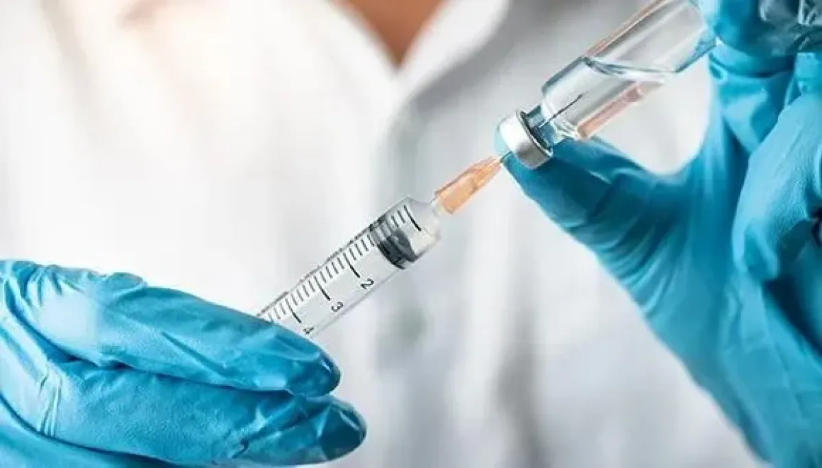
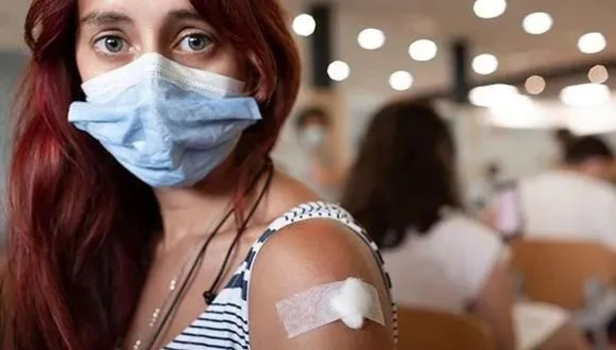
Vaccines of dietary supplements can help protect us against Corona and winter illnesses.

Since the outbreak of the Covid-19 pandemic, humanity has led an arms race against mutations, variants, and extensive contagion, in order to minimize damage to human life and the economy. Currently the battle against the virus relies mainly on RNA-based vaccines, alongside several anti-viral medications.

The Covid virus, however, changes very rapidly, and frequent updates are required to treatments and vaccines that are based on familiarizing the immune system with the virus. The same is true for flu viruses, another widespread cause of illness and death. Now, TAU researchers have shown that common dietary supplements can help protect us against the Covid virus as well as several common winter illnesses.

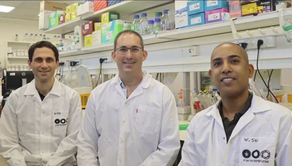
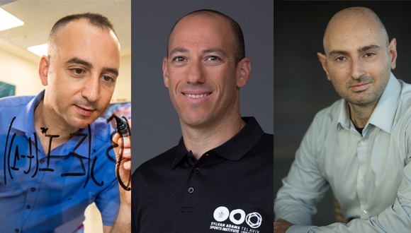
The study was led by Prof. Ehud Gazit, Prof. Eran Bacharach, and Prof. Daniel Segal of The Shmunis School of Biomedicine and Cancer Research at The George S. Wise Faculty of Life Sciences, TAU, together with PhD students Topaz Kreiser and David Zaguri and other researchers. The paper was published in Pharmaceuticals.

Preventive Supplement

Prof. Gazit, who also heads TAU’s BLAVATNIK CENTER for Drug Discovery, said: “To address the rapid changes of the virus, we decided to develop active vaccines made of safe and easily obtainable dietary supplements, that would reduce the viral load in the body and cut down contagion. We have known for years that food supplements containing zinc can enhance immunity to severe, viral, and chronic infections and their potentially grave consequences.”

The researchers found that the consumption of zinc alone achieves a relatively low cellular content. To enhance the effect, they combined the zinc with flavonoids – polyphenolic compounds found in many fruits and vegetables. They also added copper – in order to prevent an ionic imbalance and improve the treatment’s effectiveness.

“The interesting aspect is the treatment’s potential flexibility,” explains Prof. Bacharach. “We found that a combination of several flavonoids with zinc helps protect cells against a wide range of RNA viruses. We believe that the product can serve as a supplementary treatment to enhance the effect of existing anti-viral vaccines and medications.”

Prof. Segal adds: “Advanced lab tests, including PCR, have shown that the new vaccines we developed did, in fact, reduce the viral load. We found a 50-95% decrease in the genomic replication of various groups of RNA viruses, including Covid-19, the flu virus, and others. These results are very promising, possibly enabling the development of an orally administered biological shelf treatment.”

So far, all experiments were conducted in vitro in the lab, but the researchers are optimistic as to the study’s practical potential. Soon they hope to launch a series of clinical trials in humans, ultimately leading to an effective treatment accessible to everyone.


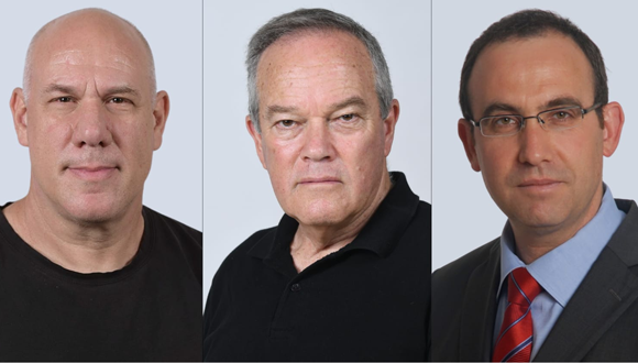
The Research Team (left to right): Professors Eran Bacharach, Daniel Segal and Ehud Gazit

Is the Vaccine Safe? Consult the Smart Sensor!

Innovative technology will prevent the reliance on self-reports for future vaccines, using smart sensors to ensure vaccine safety.

In most methods used today, clinical trials designed to evaluate the safety of a new drug or vaccine employ self-report questionnaires, asking participants how they feel before and after receiving the treatment. A new study from Tel Aviv University enables developers, for the first time in the world, to determine vaccine safety via smart sensors that measure objective physiological parameters. According to the researchers, most clinical trials testing the safety of new vaccines, including COVID-19 vaccines, rely on participants’ subjective reports, which can lead to biased results. In contrast, objective physiological data, obtained through sensors attached to the body, is clear and unambiguous.

The study was led by Dr. Yftach Gepner of the Department of Epidemiology and Preventive Medicine at TAU’s Sackler Faculty of Medicine, together with Dr. Dan Yamin and Dr. Erez Shmueli from TAU’s The Iby and Aladar Fleischman Faculty of Engineering. The paper was published in Communications Medicine, a journal from the Nature portfolio. 

The End of an Era?

Researchers from Tel Aviv University demonstrated that smart sensors can be used to test new vaccines. The current study was conducted when many Israelis received their second dose of the COVID-19 vaccine. The researchers equipped volunteers with innovative, FDA-approved sensors developed by the Israeli company Biobeat. Attached to their chests, these sensors measured physiological reactions from one day before to three days after receiving the vaccine.

The innovative sensors monitored 13 physiological parameters, such as: heart rate, breathing rate, saturation (blood oxygen levels), heartbeat volume, temperature, cardiac output, and blood pressure. The surprising results:

  • A significant discrepancy was found between subjective self-reports about side effects and actual measurements. That is, in nearly all objective measures, significant changes were identified after vaccination, even for subjects who reported having no reaction at all.
  • In addition, the study found that side effects escalate over the first 48 hours, and then parameters return to the level measured before vaccination. In other words: a direct assessment of the vaccine’s safety identified physiological reactions during the first 48 hours, with levels re-stabilizing afterwards.

“The message from our study is clear,” says Dr. Gepner. “In 2022 the time has come to conduct continual, sensitive, objective testing of the safety of new vaccines and therapies. There is no reason to rely on self-reports or wait for the occurrence of rare side effects like myocarditis, an inflammation of the heart muscle, which occurs in one of 10,000 patients. Preliminary signs that predict such conditions can be detected with advanced sensors, identifying normal vs. extreme alterations in physiological parameters and any risk of inflammation. Today, trial participants are invited to the clinic for blood pressure testing, but often their blood pressure rises just because the situation is stressful. Continual monitoring at home solves these problems with simple, convenient, inexpensive, and accurate means. This is the kind of medicine we should strive for in 2022.”


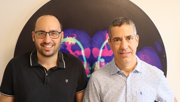
The Research Team (from left to right): Dr. Dan Yamin, Dr. Yftach Gepner and Dr. Erez Shmueli

TAU Nanodrug Enables 2-in-1 Attack on Cancer

Innovation pinpoints hard-to-treat cancers and amplifies their responsiveness to treatments.

To overcome the resistance of certain cancers to different types of treatments, Tel Aviv University researchers developed a nanodrug technology that simultaneously delivers two therapies to attack malignancy with precision. The approach lays the groundwork for cancer treatments that can work faster and with fewer side effects than existing methods.

“In our system, a single nanoparticle is capable of operating in two different arenas,” explains lead investigator Prof. Dan Peer, TAU’s Vice President of R&D, who heads the Laboratory of Precision Nanomedicine at the Shmunis School of Biomedicine and Cancer Research, The George S. Wise Faculty of Life Sciences. “It increases the receptiveness of cancer cells resistant to chemotherapy, while also reinvigorating immune cells and increasing their sensitivity to cancer cells. Thus, with one precisely targeted nanoparticle we provide two different treatments, at very different sites.”

Chemo-immunotherapy, which combines chemotherapy with immunotherapy, is considered the most advanced standard of care for various types of cancer. While chemotherapy destroys cancer cells, immunotherapy encourages the immune system to identify and attack diseased cells while sparing healthy cells critical to recovery. However, many patients fail to respond to chemo-immunotherapy, indicating the need for treatments that target cancer with greater accuracy.


Potential to Heal

In a study, Peer’s team showed how a single minescule particle, called a lipid nanoparticle, acts as a molecular precision-guided missile to deliver the two-in-one medicine directly to cancer cells. The medicine, an advanced RNA (ribonucleic acid)-based compound, alters how cancer cells function to make them identifiable for chemotherapy and immunotherapy for obliteration. 

“This is only an initial study, but it has enormous potential for positive change in the ongoing fight against cancer,” says Peer, who is a global pioneer in the field of RNA medicines. Dr. Seok- Beom Yong, a post-doctoral researcher at Peer’s lab, co-led the study. Their team tested the system in lab models for metastasized melanoma, the most aggressive type of skin cancer which spreads to other parts of the body, along with a local solid tumor confined to a single organ.

“In both populations we observed positive effects of our drug delivery system,” adds Peer, who is a member of the Roman Abramovich Center for Nanoscience and Nanotechnology at TAU. The results were published in the scientific journal Advanced Materials.

Targeted Treatment

The new development by Peer’s team builds on a recent discovery by international scientists that sheds light on how cancer evades common treatments. The discovery demonstrates how an enzyme called HO1 is used by cancer cells to both resist chemotherapy and conceal themselves from the immune system. Silencing HO1 in tumors is thus considered an optimal strategy in clinical research, but so far, all attempts to silence the enzyme led to severe side effects.

“Existing methods for silencing HO1 resemble using an F-16 fighter jet to blast a tiny ant,” says Peer. “Our new nanodrug knows how to precisely target the cancer cells, silence the enzyme, and expose the tumor to chemotherapy, without causing any damage to surrounding healthy cells. Afterwards, the same nanoparticle goes on to reprogram T-cells in the immune system to restore their ability to recognize cancer as a foreign body and attack it.”

The study was funded by an ERC grant from the European Union and a research fellowship from the South Korean government.

TAU Welcomes Ukrainian Emergency Fellowship Students

Some “need time to unfreeze”, as they begin their studies on campus.

Tel Aviv University officially welcomed seven Ukrainian graduate students, who arrived within the framework of the Emergency Fellowship Fund recently announced by the University in light of Russia’s invasion of Ukraine and the ensuing refugee crisis.

The all-women group of students hail from different cities in Ukraine, stretching from Lviv and Kyiv to Mariupol and Mikolaiv, and will continue their studies in law, medicine, psychology, music and linguistics. 

“You are very much wanted here at TAU,” President Ariel Porat told the students at the introductory meeting, expressing hope that despite the unfortunate circumstances students will find “a home away from home” at the University that will enrich their academic and personal lives. 

Constant Worry

Most of the students left their families behind in Ukraine, and worry about their wellbeing around the clock. “I managed to speak to my family yesterday, but today the connection was severed and I was unable to reach them,” says Alisa, a graduate student in law, who will be studying Crisis Management at TAU. She comes from a small town near Mariupol, in Eastern Ukraine, which has suffered some of the heaviest blows in the fighting. Alisa heard about the Fellowship through her academic advisor, as did most of the other students. 

Marina, another law student, was enrolled at the Ukrainian State Pedagogical University in Kropyvnytskyi, a central town which she says is pretty safe for now. The University premises, however, have been converted into living quarters for people escaping from more dangerous areas. Lectures are only taking place online and are highly irregular. “I was supposed to graduate in June,” she tells us, “but for now, I’m just happy to be able to continue my studies here at TAU.”

Kateryna from Kyiv studies psychology, and left immediate family members in Ukraine. “This is my first time in Israel and I know nothing about the local culture, but I’m very curious to learn,” she says, adding that the adjustment process helps her endure the constant concern about her family’s wellbeing. 

“We need some time to ‘unfreeze’, before we can start to take in and appreciate our surroundings,” adds Alisa. 

Here to Help

The students are being offered counseling and psychological services by TAU International, which has been taking full care of them since their arrival in Israel. “In light of the humanitarian situation in Ukraine, we are making a great effort to ensure that the Ukrainian students enjoy their campus experience and have a smooth transition to living in Tel Aviv, and that all their immediate needs are met,” says Michal Linder Zarankin, the School’s International Projects Coordinator.

Their tuition and living expenses are covered by TAU’s $1 million Emergency Fellowship Fund, which was swiftly raised by the University’s donors around the world over the last few weeks. 

Five more Ukrainian students are expected to arrive next week, as well as some faculty members. 

Out of the 30,000 students studying at TAU, over 300 hold dual Israeli-Ukrainian citizenship. In addition to these, there are many Israeli TAU students of Ukrainian and Russian descent. 

Featured image: Ukrainian graduate students are welcomed by TAU’s President Ariel Porat, Prof. Milette Shamir VP International and TAU International staffers

Prof. Ehud Gazit – First Israeli to Receive Prestigious International Recognition in Chemistry

Selected as International Solvay Chair in Chemistry for 2023.

Prof. Ehud Gazit from The Shmunis School of Biomedicine and Cancer Research at The George S. Wise Faculty of Life Sciences and The Department of Materials Science and Engineering at The Iby and Aladar Fleischman Faculty of Engineering, was selected as the International Solvay Chair in Chemistry for 2023. Prof. Gazit, who also heads TAU’s Blavatnik Center for Drug Discovery, is the first Israeli to receive this annually awarded honor and the first scientist to be appointed to the position outside of the United States and Europe. 

 Joining 15 Other World Top Scientists

The Solvay International Institute was founded in Belgium about a century ago and is designed to develop and support creative and groundbreaking research in physics, chemistry and related fields, in order to increase and deepen the understanding of natural phenomena. The Institute organizes annual conferences on physics and chemistry, as well as international workshops for the training of doctoral students and selected topics. 

As part of Gazit’s new appointment, he will spend a month or two in Brussels, the capital of Belgium, during which he will give lectures on his field of research. The prestigious nomination has previously been awarded to 15 of the world’s top scientists, including three Nobel laureates in chemistry, the Wolf Prize winner and laureates of other prestigious awards, all from leading institutions in the US and Europe, who are now joined by Gazit. 

Gazit is a biophysicist, biochemist and nanotechnologist. His main area of expertise is “Solid State Biology”, an innovative field of study that combines disciplines from physics, chemistry, synthetic and structural biology and materials engineering. He is a world-renowned expert in nanotechnology and biological chemistry, a highly cited researcher who has published more than 350 scientific articles and inventor of more than 100 patents.

Previously, he served as Vice President for Research and Development of the University, as the Chairman of Ramot, Tel Aviv University’s Tech Transfer Company, and as the Chief Scientist of Israel’s Ministry of Science and Technology. 

Over the years, Gazit has won a number of prestigious awards and prizes in Israel and around the world, including The Kadar Family Award for Outstanding Research, the Landau Prize in Science and Arts and the Rapaport Prize for Excellence in Biomedical Research. He is a Fellow of the Royal Society of Chemistry in the UK, a Foreign Fellow of the National Academy of Sciences in India and a Member of the European Organization for Molecular Biology.  

Gazit stated: “I thank the Solvay Institute for selecting me, a great honor and excitement for me. It is a great privilege for me to join such a prestigious and impressive list of leading researchers. Today I am reminded of the former President of Israel, Prof. Ephraim Katzir, one of Israel’s greatest scientists, and of whom I am one of his academic ‘great grandchildren’ and who organized the Solvay Institute’s Chemistry Conference about 40 years ago. Apart from the personal honor, I am happy and proud to represent Tel Aviv University and the State of Israel in this appointment.”

Minor Head Injury Leads to Chronic Post-Concussion Syndrome in 1 of 4 Children

Long term consequences of mild head injury in children are underdiagnosed, causing prolonged suffering.

A new study by Tel Aviv University, Kaplan Medical Center and Shamir Medical Center (Assaf Harofeh) found that one in four children (25.3%) who have been discharged from the emergency room after a mild head injury are misdiagnosed and continue to suffer from persistent post-concussion syndrome for many years. The syndrome includes chronic symptoms such as memory loss, psychological issues and sensitivity to light and noise, which can be misdiagnosed as symptoms of ADHD, depression or sleeping disorders. The misdiagnosis results in the children receiving treatment that is not suited to their condition, which causes them prolonged suffering.

The study was led by Prof. Shai Efrati of the Sagol Center for Hyperbaric Medicine and Research at Tel Aviv University and Shamir Medical Center (Assaf Harofeh), Dr. Uri Bella and Dr. Eli Fried of Kaplan Medical Center, and Prof. Eran Kotzer of Shamir Medical Center. The results of the study were published in the journal Scientific Reports.

The researchers examined 200 children who suffered from a head injury and who were released from the emergency room after the need for medical intervention was ruled out. They tracked the subjects for a period between six months and three years, and found that about one in four children released from the emergency room suffered from the chronic syndrome.

Long-Term Monitoring Needed

According to the researchers, the alarming findings demonstrate that changes in the approach are needed to be monitoring and treating these children. 

“Persistent post-concussion syndrome is a chronic syndrome that results from micro damage to the small blood vessels and nerves, which may appear several months after the head injury. As a result, it often gets misdiagnosed as attention deficit disorders, sleep disorders, depression, and so on,” explains Prof. Shai Efrati. 

“The purpose of an emergency room diagnosis is to determine whether the child suffers from a severe brain injury that requires immediate medical intervention,” adds Prof. Eran Kotzer, Director of the Emergency Rooms at the Shamir Medical Center.  “Unfortunately, the way most medical systems operate today, we miss long-term effects and do not continue to monitor those children who leave the emergency room without visible motor impairment.”

“Treatment for a wide range of disorders will change if we know that the cause of the new problem is a brain injury,” concludes Prof. Efrati. “Proper diagnosis of the cause is the first and most important step in providing appropriate treatment for the problem.”

Inventive Study to Develop Biological Solutions for Agriculture

TAU and ag-biotech company PlantArcBio to collaborate on development of RNAi-based products.

Genetically improved plants can be a real-life magic stick for solving global famine issues. In a first-of-its-kind study, Ramot, the Technology Transfer Company of Tel Aviv University will cooperate with ag-biotech company PlantArcBio to develop innovative RNAi-based biological solutions for agriculture.

RNAi technology enables a temporary external disruption of RNA (ribonucleic acid) molecules, diminishing the amount of Messenger RNA (mRNA), thus temporarily reducing the expression of specific genes, without modifying or genetically engineering the organism’s DNA. Externally applied RNAi molecules affect specific genes for a specific time period, as required for positive effects like crop protection and yield enhancement. 

Specifically, the research will focus on testing the joint technology’s contribution to the stability of RNAi-based products and their ability to penetrate plants and insects.

Joining Forces

The first-of-its-kind joint study will examine the efficacy of PlantArcBio‘s RNAi technology for agriculture, combined with the unique lipid-based RNA delivery technology developed by Prof. Dan Peer, TAU’s Vice President for R&D, head of the Center for Translational Medicine and a member of both the Shmunis School of Biomedicine and Cancer ResearchGeorge S. Wise Faculty of Life Sciences, and the Center for Nanoscience and Nanotechnology, and a pioneer using RNA to manipulate cells in cancer and other immune related diseases.  


Prof. Dan Peer

“We see great value in contributing to the development of RNAi-based products addressing global issues and providing an ecological and environmentally friendly solution to the global challenges of sustainability in agriculture and food security,” says Peer.

Keren Primor Cohen, CEO of Ramot, believes there is “extensive commercial potential for this combined technology” and welcomes the collaboration with PlantArcBio.

The research will be carried out both at PlantArcBio‘s Laboratories and at Prof. Dan Peer’s Laboratory of Precision NanoMedicine at Tel Aviv University. According to Dror Shalitin, Founder and CEO of PlantArcBio, the results are expected within approximately 12 months.

TAU Technology Could Prevent Repeat Heart Valve Surgery

Invention may lead to a dramatic improvement in the quality of life of many heart patients.

An international study led by a researcher from Tel Aviv University offers a novel technology that can assist many patients implanted with bioprosthetic heart valve by avoiding additional complicated replacement surgery. Today, many heart patients implanted with such valves are forced to replace it ten years later due to calcification of valve tissue. The researchers have now been able to show that by genetically engineering the biological component in the valve, immunological attack and calcification risk can be avoided – thereby offering next-generation durable bioprosthetic heart valves.

This technological development stems from the EU-funded TRANSLINK consortium consisting of 14 members from Europe, USA and Canada and led by Dr. Vered Padler-Karavani from The Shmunis School of Biomedicine and Cancer Research, The George S. Wise Faculty of Life Sciences at Tel Aviv University. In this study, the researchers investigated close to 1700 patients with ~5000 blood samples covering almost 15 years since implantation. The study was published in the prestigious journal Nature Medicine.

The researchers explain that patients can be implanted with either a mechanical heart valve that last long but requires daily administration of anticoagulants that can lead to life-threatening hemorrhages, or bioprosthetic heart valves (made from bovine, porcine or equine tissue) allowing patients to live a reasonably normal life, but those are commonly destroyed after a decade requiring replacement.

Getting Rid of Foreign Sugars

Dr. Padler-Karavani: “since bioprosthetic heart valves are made of animal tissues, we hypothesized they contain foreign non-human sugars (Neu5Gc and alpha-Gal) that are attacked by the human immune system, which then mediate the calcification that lead to structural valve deterioration. Indeed, in our research we proved that this was the reason and even suggested an implementable solution.

“We discovered that all bioprosthetic heart valve patients developed an immune response against the foreign sugars in the valves. We could clearly see an increase in antibody responses against these sugars in implanted patients, as early as one month after implantation, some lasting even two years later. We also found that some of the patients showed signs of calcification as early as two years post implantation”.


Dr. Vered Padler-Karavani

Anu Paul, a postdoc in the lab, also showed that the foreign sugars and the antibodies attacking them were found on calcified bioprosthetic heart valves explanted from patients some 10 years after implantation. Additionally, the dietary non-human sugar Neu5Gc and the antibodies against it were also found on explanted calcified native valves (the original malfunctioning human valves that needed to be replaced with a bioprosthesis). Since this sugar cannot be produced in the human body, it most likely accumulates on these valves from diet rich in red meat and dairy products where it is abundant. Therefore, it is possible that red meat diet mediates the initial need for valve replacement. The researchers also confirmed in a human-like animal model that antibodies against the foreign sugars indeed mediate calcification of tissues used for production of bioprosthetic heart valves.

Furthermore, the option to employ genetic engineering to resolve the problem was examined. For this purpose, the consortium created genetically modified porcine (pigs) that do not express the sugars foreign to humans. In Dr. Padler-Karavani’s lab the researchers found that in a human-like animal model that engineered tissue lacking the foreign sugars significantly had reduced calcification even in the presence of antibodies against the sugars and can therefore increase the durability of bioprosthetic heart valves made of such tissues.

“This study marks breakthrough technology in the field of bioprosthetic heart valves and provides deep understanding of the mechanisms leading to structural valve deterioration. These findings can lead to a dramatic improvement in the quality of life of many heart patients. Now, it would be interesting to study whether vegetarians or people who consume only small amounts of red meat and dairy have lower probability of heart valve calcification, and if this could perhaps be associated with low levels of antibodies against these foreign sugars. In the future, it may also be possible to devise a modified diet to reduce the risk or to actually produce biological valves from the tissues of engineered animals that do not contain the sugars at all”, Dr. Padler-Karavani summarizes.

Israeli Breakthrough in Treating PTSD

World first: TAU-led team shows success of oxygen therapy in alleviating symptoms of PTSD in military veterans.

Researchers from Tel Aviv University and Israel’s Shamir Medical Center were able to successfully relieve the symptoms of post traumatic stress disorder (PTSD) in military combat veterans using a new protocols of hyperbaric oxygen therapy (HBOT). In a controlled clinical trial involving Israeli army veterans who suffered from treatment-resistant PTSD, the approach demonstrated significant improvement in all classes of symptoms.

According to the World Health Organization (WHO), almost 4% of the global population, and 30% of all combat soldiers, develop PTSD. 

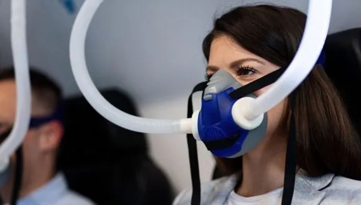
Hyperbaric medicine involves treatments in a pressurized chamber where atmospheric pressure is higher than sea-level pressure and the air is rich with oxygen. Considered a safe form of treatment, hyperbaric oxygen therapy is already used for a range of medical conditions. Evidence gathered in recent years indicates that special hyperbaric protocols can improve the supply of oxygen to the brain, thereby enhancing the generation of new blood vessels and neurons. It must be noted that HBOT treatments require the evaluation and supervision of qualified physicians. Moreover, for medical indications it should be given using a certified chamber with appropriate quality assurance using the exact studied treatment protocols. 

The breakthrough research was led by Prof. Shai Efrati, Dr. Keren Doenyas-Barak, and Dr. Amir Hadanny of Tel Aviv University’s Sackler Faculty of Medicine and Sagol School of Neuroscience in cooperation with Shamir Medical Center. The team also included Dr. Ilan Kutz, Dr. Merav Catalogna, Dr. Efrat Sasson, Gabriela Levi and Yarden Shechter of Shamir Medical Center.

Unloading Pain for a Better Future

The study included 35 combat veterans of the Israel Defense Forces (IDF) who suffered from PTSD that was resistant to both psychiatric medications and psychotherapy. 

“The veterans were divided into two groups: one group received hyperbaric oxygen therapy while the other served as a control group,” explains Dr. Keren Doenyas-Barak of Shamir Medical Center. “Following a protocol of 60 treatments improvement was demonstrated in all PTSD symptoms, including hyper-arousal, avoidance, and depression. Moreover, both functional and structural improvement was observed in the non-healing brain wounds that characterize PTSD. We believe that in most patients, improvements will be preserved for years after the completion of the treatment.”

“This study gives real hope to PTSD sufferers. For the first time in years the study’s participants, most of whom had suffered from severe PTSD, were able to leave the horrors behind and look forward to a better future.”

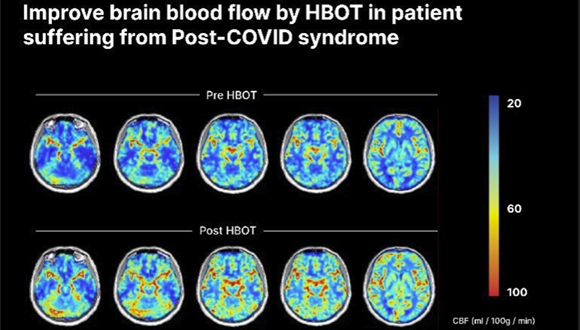
Illustration: Clinical example of functional brain imaging by fMRI.  The reduced brain activity in the frontal lobes of the brain (responsible among others for emotional regulation and executive functions) and in hippocampus (responsible for memories functions) is improved after Hyperbaric Oxygen Therapy (HBOT).

Emotional Trauma Can Cause Physical Damage

“Today we understand that treatment-resistant PTSD is caused by a biological wound in brain tissues, which obstructs attempts at psychological and psychiatric treatments,” explains TAU Prof. Shai Efrati. “With the new hyperbaric oxygen therapy protocols, we can activate mechanisms that repair the wounded brain tissue. The treatment induces reactivation and proliferation of stem cells, as well as generation of new blood vessels and increased brain activity, ultimately restoring the functionality of the wounded tissues. Our study paves the way to a better understanding of the connection between mind and body.”

“Our results indicate that exposure to severe emotional trauma can cause organic damage to the brain,” says Prof. Efrati. “We also demonstrate for the first time that direct biological treatment of brain tissues can serve as a tool for helping PTSD patients. Moreover, our findings may be most significant for diagnosis. To date, no effective diagnostic method has been developed and diagnosis of PTSD is still based on personal reports which are necessarily subjective – leading to many clashes between the suffering veterans and the authorities responsible for treating them. Think of a person who comes to the emergency room with chest pains. The pain might be caused by either a panic attack or a heart attack, and without objective EKG and blood tests, the doctors might miss a heart attack. At present we are conducting continuing research in order to identify the biological fingerprint of PTSD, which can ultimately enable the development of innovative objective diagnostic tools.”


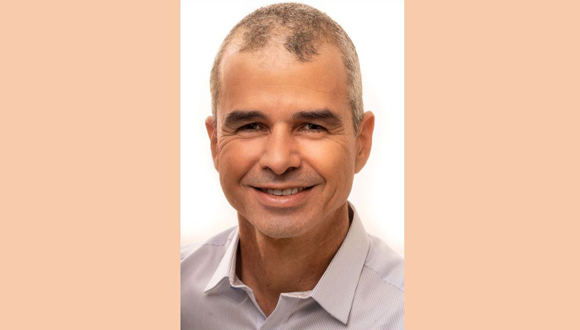
Prof. Shai Efrati

Prof. Efrati is an Associate Professor at TAU and director of the Sagol Center for Hyperbaric Medicine and Research at Shamir Medical Center. He is also the co-founder and Chair of the Medical Advisory Board at Aviv Scientific LTD, a company that applies the hyperbaric oxygen therapy protocols developed from his team’s research to enhance the brain and body performance of aging adults. 

Discovery May Enable Early Diagnosis of Alzheimer’s

Can your sleep predict your chance of developing the disease?

A new study at Tel Aviv University revealed abnormal brain activity that precedes the onset of Alzheimer’s first symptoms by many years: increased activity in the hippocampus, a region of the brain which plays a key role in memory processes, during anesthesia and sleep, resulting from failure in the mechanism that stabilizes the neural network. The researchers believe that the discovery of this abnormal activity during specific brain states may enable early diagnosis of Alzheimer’s, eventually leading to a more effective treatment of a disease that still lacks effective therapies.

The study was published in the prestigious scientific journal Cell Reports, and led by Prof. Inna Slutsky and doctoral students Daniel Zarhin and Refaela Atsmon from the Sackler Faculty of Medicine and the Sagol School of Neuroscience at Tel Aviv University.

According to Prof. Inna Slutsky, innovative imaging technologies developed in recent years have revealed that amyloid deposits, a hallmark of Alzheimer’s disease pathology, are formed in patients’ brains as early as 10-20 years before the onset of typical symptoms such as memory impairment and cognitive decline. Unfortunately, most efforts to treat Alzheimer’s disease have failed. She believes that if we could detect the disease at the pre-symptomatic stage, and keep it in a dormant phase for many years, this would be a tremendous achievement in the field. Identifying a signature of aberrant brain activity in the pre-symptomatic stage of Alzheimer’s and understanding the mechanisms underlying its development she says may be a key to effective treatment.

Additional participants in the study include: Dr. Antonella Ruggiero, Halit Baeloha, Shiri Shoob, Oded Scharf, Leore Heim, Nadav Buchbinder, Ortal Shinikamin, Dr. Ilana Shapira, Dr. Boaz Styr, and Dr. Gabriella Braun, all from Prof. Slutsky’s laboratory. Collaborations with the laboratory teams of Prof. Yaniv Ziv of the Weizmann Institute, and Prof. Yuval Nir of TAU were essential for the project. Prof. Tamar Geiger, Dr. Michal Harel, and Dr. Anton Sheinin of Tel Aviv University, as well as researchers from Japan, also contributed to the study.

The researchers used animal models for Alzheimer’s, focusing on the hippocampal region of the brain, which is known to be impaired in Alzheimer’s patients. At first, they measured cell activity in the hippocampus when the model animal was awake and active. For this, they used advanced methods that measure brain activity at a resolution of single neurons.

High Neuronal Activity – Also During Sleep

“It is known that neuronal activity of the hippocampus decreases during sleep in healthy animals,” explains Refaela Atsmon. However, when she examined model animals in early stages of Alzheimer’s, she found that their hippocampal activity remained high even during sleep. This is due to a failure in the physiological regulation, which she says has never before been observed in the context of Alzheimer’s disease.

Daniel Zarhin found similar dysregulation in model animals under anesthesia: neuronal activity did not decline, the neurons operated in a manner that was too synchronized, and a pathological electrical pattern was formed, similar to ‘quiet’ seizures in epileptic patients.

The researchers found that brain states that block responses to the environment – such as sleep and anesthesia – expose abnormal activity which remains hidden when the animal is awake, and this happens before the symptoms of Alzheimer’s disease are observed.

Prof. Slutsky’s explains that even though this abnormal activity can be detected during sleep, it is much more frequent under anesthesia. Therefore, she says, it would be important to test whether short anesthesia can be used for early diagnosis of Alzheimer’s disease.

Defective Stabilizing Mechanisms

The researchers proceeded to ask what causes the abnormality. To this end, they relied on findings from previous studies from Prof. Slutsky’s laboratory and other researchers on homeostasis of neural networks: each neural circuit has a set point of activity, maintained by numerous stabilizing mechanisms. These mechanisms are activated when the balance is disturbed, restoring neuronal activity to its original set point.  

Is a disruption of these mechanisms the main cause of deviant brain activity during sleep and anesthesia in Alzheimer’s disease animal models? To test this, Dr. Antonella Ruggiero examined the effect of various anesthetics on neurons grown on a chip. She found that they lower the set point of neuronal activity. While in healthy neural networks this activity remained low over time, in neural networks expressing genetic Alzheimer’s mutations, the lowered set point recovered quickly, despite the presence of anesthetics.

The researchers now sought to examine a potential drug for the impaired regulatory mechanism. According to Prof. Slutsky, the instability in neuronal activity found in the study is known from epilepsy. In a previous study Prof. Slutsky’s team discovered that an existing drug for multiple sclerosis may help epilepsy patients by activating a homeostatic mechanism that lowers the set point of neural activity. Doctoral student Shiri Shoob examined the effect of the drug on hippocampal activity in the animal model for Alzheimer’s and found that also in this case the drug stabilizes activity and reduces pathological activity observed during anesthesia.

Proceeding Towards Clinical Trials

“The results of our study may help early diagnosis of Alzheimer’s, and even provide a solution for instability of neuronal activity in Alzheimer’s disease,” says Prof. Slutsky. Firstly, we discovered that anesthesia and sleep states expose pathological brain activity in the early stages of Alzheimer’s disease, before the onset of cognitive decline. We also proposed the cause of the pathological activity – failure of a very basic homeostatic mechanism that stabilizes electrical activity in brain circuits. Lastly, we showed that a known medication for multiple sclerosis suppresses this type of anesthesia-induced aberrant brain activity,” she concludes.

The researchers now plan to collaborate with medical centers in Israel and worldwide to test whether the mechanisms discovered in animal models can also be identified in patients with early-stage Alzheimer’s disease. For this purpose, they propose to incorporate EEG monitoring into surgical procedures, to measure brain activity of patients under anesthesia. They hope that their findings will promote early diagnosis and drug development for the most common form of late-onset dementia.

Featured image: The Research Team (from left to right): Prof. Inna Slutsky, Daniel Zarhin and Refaela Atsmon (Photo: Dr. Tal Laviv)

Victoria

Tok Corporate Centre, Level 1,
459 Toorak Road, Toorak VIC 3142
Phone: +61 3 9296 2065
Email: [email protected]

New South Wales

Level 22, Westfield Tower 2, 101 Grafton Street, Bondi Junction NSW 2022
Phone: +61 418 465 556
Email: [email protected]

Western Australia

P O Box 36, Claremont,
WA  6010
Phone: :+61 411 223 550
Email: [email protected]