
Tag: Neuroscience

Why Do Bats Fly Into Walls?

A sensory misperception – like people bumping into a glass wall

Why do bats fly into walls, even though they can hear them? Researchers at Tel Aviv University conducted an experiment in which they released dozens of bats in a corridor blocked by objects of different sizes, made of different materials. To their surprise, the researchers discovered that the bats collided with large sponge walls (that produce a weak echo) as if they did not exist. The bats’ behavior suggested that they did this even though they had detected the wall with their sonar system, indicating that the collision did not result from a sensory limitation, but rather from an acoustic misperception. The researchers hypothesize that the unnatural combination of a large object (wall) and a weak echo disrupts the bats’ sensory perception and causes them to ignore the obstacle (much like people who bump into transparent walls).

The study was led by Dr. Sasha Danilovich then a PhD student in the lab of Prof. Yossi Yovel, Head of the Sagol School for Neuroscience and faculty member at the School of Zoology at the George S. Wise Faculty of Life Sciences. Other participants included Dr. Arian Bonman and students Gal Shalev and Aya Goldstein of the Sensory Perception and Cognition Laboratory at the School of Zoology and the Sagol School of Neuroscience. The paper was published in PNAS.

At the next stage of the experiment, the researchers methodically changed the features of the echoing objects along the corridor in terms of size, texture and echo intensity. They concluded that the bats’ acoustic perception depends on a coherent, typical correlation of the dimensions with objects in nature. For example: large object- strong echo; small object – weak echo.

“Bats excel in acoustic perception. They are able to detect objects as tiny as mosquitoes, using sound waves,” explains Prof. Yovel. “Using echolocation they can calculate the 3-dimensional location of both small and large objects, perceiving their shape, size and texture. To this end a bat’s brain processes various acoustic dimensions from the echoes returning from the object (such as frequency, spectrum and intensity). This perception is based on several senses that combine many different dimensions, such as color and shape.”

In addition, the researchers at TAU discovered that bats are not born with this ability. Repeating the experiment with young bats they found that they do not fly into walls.  The study also found that adult bats can quickly learn the new correlations among the dimensions.

“By presenting the bats with objects whose acoustic dimensions are not coherent, we were able to mislead them, creating a misconception that caused them to repeatedly try to fly through a wall, even though they had identified it with their sonar. The experiment gives us a peek into how the world is perceived by these creatures, whose senses are so unique and different from ours,” says Sasha Danilovich.

TAU Prof. Wins Schmidt Science Polymath Award

Prof. Oded Rechavi one of first winners of prestigious prize, which is defined as “an experiment in extreme curiosity-driven innovation”

A great honor for Israeli science: Schmidt Futures, a philanthropic initiative founded by Eric and Wendy Schmidt,  has decided to establish a new $2.5m award entitled Polymaths, for researchers exhibiting rare interdisciplinarity. Only two scientists have been chosen to receive the first Polymaths Award: Prof. Jeff Gore of MIT and Prof. Oded Rechavi of the Neurobiology Department at the George S. Wise Faculty of Life Sciences and the Sagol School of Neuroscience at Tel Aviv University. Each of the two scientists will receive an annual unrestricted grant of $500,000 for five years, to pursue any direction of research. “I am proud to have been chosen and excited about the opportunity to open new fields of research,” says Prof. Rechavi. “Typically scientists receive funds for research projects that are already underway. The Polymaths Award is different. They tell you: ‘Here are the resources. Do something completely new, take risks. Investigate wild ideas you never would have dreamed of proposing to other research foundations.'”

The Schmidt Science Polymath program is an initiative created under Schmidt Futures, which finds exceptional people and helps them achieve more for others by applying advanced science and technology thoughtfully and by working together across fields. The program aims to provide outstandingly interdisciplinary researchers with the means to expand their research even further. In the future, a prestigious network of the award’s laureates will be established. “An experiment in extreme curiosity-driven innovation,” proclaims the program. “Instead of focusing on specific research ideas, the goal for the program is to bet on people, their special talents, and their teams.” 

The laboratory of the first Polymath Award laureate, Prof. Oded Rechavi, excels in promoting interdisciplinary research. In recent years Prof. Rechavi has studied a very vast range of topics, achieving scientific breakthroughs in fields that are not necessarily connected to one another. Thus, for example, Rechavi discovered a mechanism enabling transgenerational inheritance of parental responses, showing for the first time that small RNAs are inherited alongside DNA, and deciphering the laws of epigenetic heredity. In another study, Rechavi and his team assisted in decoding the Dead Sea Scrolls through the DNA of the parchments on which they were written, shedding more light on the history of the late Second Temple period. Rechavi also explored the neuronal basis of irrationality, finding a simple law for altering the nervous system of worms so that they become less or more rational. In a completely different area, Rechavi’s group genetically engineered parasites to turn them into protein-secreting machines enabling repair of genetic diseases of the nervous system.

Prof. Oded Rechavi. Photo: Yehonatan Zur.

How the parents’ environment impacts the lives of their offspring

Three rules that dictate transgenerational epigenetic inheritance in worms – independently of changes in DNA sequences.

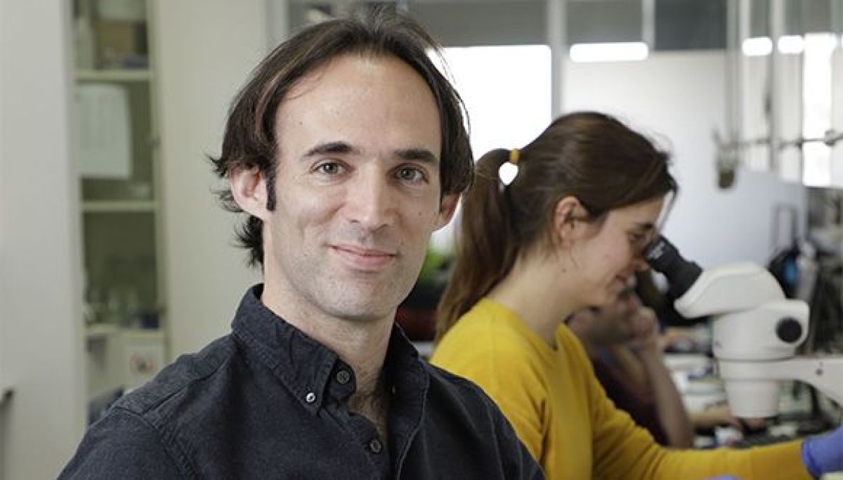
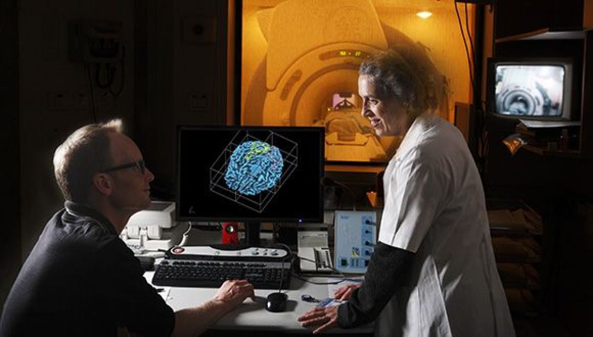
Researchers at Tel Aviv University have discovered three rules that dictate epigenetic inheritance – meaning transgenerational inheritance through means other than changes in DNA sequences. Published today in the leading scientific journal Cell, the study was led by Prof. Oded Rechavi and his research student Dr. Leah Houri-Zeevi of the Department of Neurobiology at the Faculty of Life Sciences and the Sagol School of Neuroscience at Tel Aviv University.

Most experiences we acquire in our lifetime will not be passed on to our descendants. For example, our workout in the gym today will not make our children stronger. However, studies conducted in recent years on epigenetic inheritance in worms challenge our traditional concepts regarding the limits of inheritance and evolution, indicating that some acquired traits are in fact passed on to subsequent generations. Prof. Rechavi explains: “Epigenetic inheritance of responses to the environment occurs independently of changes to the DNA sequence, through other inherited molecules. In many organisms, responses to environmental changes, such as stress, involve small RNA molecules that silence or block the expression of certain genes.” In recent years, research on C. elegans worms – an important and widely used model animal – has shown that small RNA molecules can be transmitted to subsequent generations, thereby passing on certain traits.

In previous research projects, Prof. Rechavi discovered that worms transfer to their offspring small RNA molecules containing information on the parent’s environment, such as viral infections, nutrition, and even brain activity, thereby contributing to the survival of subsequent generations. In the current study, Prof. Rechavi and his team tried to understand whether transgenerational epigenetic inheritance via small RNA molecules is governed by specific rules, or alternately, occurs passively and randomly.

According to Prof. Rechavi, “C. elegans is the preferred model organism for research on transgenerational epigenetic inheritance, for several  reasons: Its generation time is three and a half days, allowing us to study many generations in a short period of time; every worm produces hundreds of descendants, providing strong statistical validity; environmental exposure can be fully controlled; and each worm fertilizes itself, so that differences in DNA are almost completely neutralized.”

Dr. Houri-Zeevi explains that “Many laboratories have noted that at the level of a population epigenetic inheritance through small RNA endures for about three to five generations in worms. In a previous study, we discovered a mechanism that controls the duration of the inheritance, proving, in effect, that this type of inheritance is a regulated process. But still the question remains: Why are some worms strongly affected by their ancestor’s environmental responses, while others do not inherit the epigenetic effect at all – despite the fact that all offspring are almost identical genetically. This partial inheritance has been known for some time, but how epigenetic material is distributed among the offspring remained a mystery. We wanted to find out whether there was any pattern in the inheritance, that might explain and allow us to predict who would inherit the epigenetic features – and for how long.”

The researchers used a genetically engineered worm carrying a gene that produces a fluorescent protein – making the worm itself glow under fluorescent light. The researchers then initiated a heritable small RNA silencing response against the fluorescent gene and observed which descendants had inherited the silencing response and stopped glowing, and which descendants ‘forgot’ the parental response and started expressing the fluorescent gene once again after several generations. Dr. Houri-Zeevi repeated this process over and over again, in an attempt to understand the rules governing the epigenetic effect. Altogether she examined dozens of worms lineages, including more than 20,000 individual worms. But the most challenging part, according to Prof. Rechavi, was deciphering the different inheritance patterns and understanding the rules behind them.

Ultimately, through in-depth investigation of the inheritance mechanism, the researchers discovered three laws that can explain and even enable the prediction of who inherits the epigenetic information:

  • First law: Inheritance is uniform in worms descending from the same mother – namely worms of the same lineage. The researchers were surprised to learn that differences in inheritance observed in previous studies were in fact ‘concealed’ due to the method of examining whole worm populations rather than distinct lineages.
  • Second law: Inheritance is very different in worms derived from different mothers, even though the mothers themselves are supposedly identical, because the worm fertilizes itself. The researchers characterized the mechanism that creates the differences between mothers who are genetically identical and found that differences between descendants stem from varying ‘internal states’ randomly adopted by the mothers. Essentially, the mother’s internal state, the level of activity of the inheritance mechanism in each mother, determines the duration of inheritance, and thus the fate of subsequent generations.
  • Third law: The longer the duration of the epigenetic inheritance – namely, the greater the number of generations in a specific lineage who inherits the trait – the greater the probability that it will continue on to the next generation as well, “in something like transgenerational momentum, resembling the ‘Hot Hand’ rule in basketball.”

According to Prof. Rechavi, we do not yet know whether the exact same transgenerational epigenetic inheritance mechanism exists in humans as well: “We hope that the mechanism we have discovered exists in other organisms as well, but we’ll just have to be patient. We must remember that genetic research also began with Friar Gregor Mendel’s observations in peas, and today we use Mendel’s laws to predict whether our children will have smooth or curly hair.”

“The idea of acquired traits passed on to descendants is as old as it is outrageous. Even before Darwin and Lamarck, the ancient Greeks argued about it, and it seems to be incompatible with genetic inheritance through DNA,” adds Prof. Rechavi. “The worms changed the rules by showing us that inheritance outside the genetic sequence does exist, via small RNA molecules, enabling parents to prepare their offspring for the difficulties they have encountered in their lifetime. From one study to the next we shed light on the molecular mechanisms and mysterious dynamics of epigenetic inheritance, with the present study providing laws and introducing some ‘order into the chaos’.”
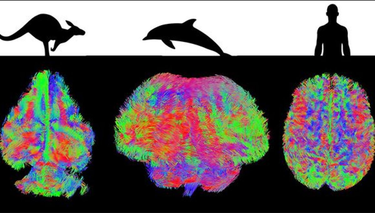
Does our Brain like risk?

A new study attempts to find out whether our brains are prone to over caution or to underestimating risk

A new Tel Aviv University study examined the brain’s reactions in conditions of uncertainty and stressful conflict in an environment of risks and opportunities. The researchers identified the areas of the brain responsible for the delicate balance between desiring gain and avoiding potential loss along the way.

The study was led by Tel Aviv University researchers Prof. Talma Hendler, Prof. Itzhak Fried, Dr. Tomer Gazit, and Dr. Tal Gonen from the Sackler Faculty of Medicine, the School of Psychological Sciences and the Sagol School of Neuroscience, along with researchers from the the Tel Aviv Sourasky Medical Center (Ichilov) and the University of California, Los Angeles School of Medicine. The study was published in July 2020 in the prestigious journal Nature Communications.

Prof. Hendler explains that in order to detect reactions in the depths of the brain, the study was performed among a unique population of epilepsy patients who had electrodes inserted into their brains for testing prior to surgery to remove the area of the brain causing epileptic seizures. Patients were asked to play a computer game that included risks and opportunities, and the electrodes allowed the researchers to record, with a high level of accuracy, neural activity in different areas of the brain associated with decision-making, emotion and memory.

Your brain suggests – play it safe

Throughout the game, the researchers recorded the electrical activity in the subjects’ nerve cells immediately after they won or lost money. The subjects were asked to try to collect coins while taking the risk of losing money from their pool. It was found that the neurons in the area of ​​the inner prefrontal cortex responded much more to loss (punishment) than to the gaining (reward) of coins.

Moreover, the researchers found that the avoidance of risk-taking in the players’ next move was affected mainly by post-loss activity in the area of the hippocampus, which is associated with learning and memory, but also with anxiety. This finding demonstrates the close relationship between memory processes and decision-making when risk is present (stressful situations). That is, the loss is encoded in the hippocampus (the region of the brain associated with ​​memory), and the participant operating in a high-risk stressful situation preferred to be cautious and avoid winning the coins (forfeiting the gain).

The experience of winning, however, was not encoded in the memory in a way that influenced the choice of future behavior in conditions of uncertainty. An interesting point is that this phenomenon was found only when the subject was the once influencing the result of the game, and only in the presence of a high risk in the next move, which indicates a possible connection to anxiety.

Prof. Hendler summarizes: “Throughout life, we ​​learn to balance the fear of risking loss with the pursuit of profit, and we learn what is a reasonable risk to take in relation to the gain based on previous experiences. The balance between these two tendencies is a personality trait but is also affected by stress (like the current pandemic). A disorder in this trait increase sensitivity to stress and can cause non-adaptive behavior such as a high propensity for risk-taking or excessive avoidance.

“Our research shows for the first time how the human brain is affected by the experience of failure or loss when it is our responsibility, and how this inclination produces avoidance behavior under particularly stressful uncertainty. An understanding of the neural mechanism involved may guide future neuropsychiatric therapies for disorders featuring excessive avoidance, such as depression, anxiety, and PTSD, or disorders associated with excessive risk-taking, such as addiction and mania.”

Featured image: Prof. Talma Hendler

Tel Aviv University Scientists Successfully Reduce Metastatic Spread Following Tumor Removal Surgery

A Study Performed in Colorectal Cancer Patients Found that Implementing a Stress-Inflammatory Response Reducing Treatment During Surgery Could Lead to a Decrease in Metastatic Risk

A research group from Tel Aviv University successfully reduced metastatic spread following tumor removal surgery in colorectal cancer patients. Using a short medication treatment around the time of the surgery, the researchers were able to reduce body stress responses and physiological inflammation during this critical period, thus preventing the development of metastases in the years following the surgery. The study, which was recently published in “Cancer”, was led by Prof. Shamgar Ben-Eliyahu from TAU School of Psychological Sciences and Sagol School of Neuroscience, and Prof. Oded Zmora from Shamir (Assaf Harofeh) Medical Center. During the study, which lasted 3 years, the researchers have monitored 34 patients, who received treatment surrounding a colorectal tumor removal surgery. During the pre- and post-surgical period, the patients were administered two safe and known drugs: Propranolol (Deralin), an anti-anxiety and blood pressure reducing drug, and Etodolac (Etopan), an anti-inflammatory analgesic. The drugs were only administered to the patients for 20 days – starting from 5 days prior to surgery, and until two weeks after – with half of the patients receiving a placebo treatment, as a control group. The results are highly promising: while only 12.5% (2 out of 16) of patients receiving the drugs treatment exhibited metastatic disease, in the control group (which did not receive the treatment) the rate of metastases development was found to be 33% (6 out of 18 patients), which is the known rate for colorectal cancer patients. Prof. Ben-Eliyahu says that he is highly satisfied with these data, but also states that “despite the impressive results, this treatment must be examined again, in a much larger number of patients, in order to test whether it is, in fact, life-saving”. According to Prof. Ben-Eliyahu, the study of molecular markers in the cancerous tissue excised from the patients showed that the treatment with the medications has led to a reduction in the metastatic potential of the tumor and potentially the residual cancer cells. In addition, the drugs triggered some beneficial alterations in infiltrating tumor leukocytes (patients’ white blood cells) number and type – which are also markers indicating a reduced chance of disease recurrence. Prof. Ben-Eliyahu explains: “When the body is in a state of stress, whether physiological (from surgery) or psychological, this causes a release of high amounts of two types of hormones, prostaglandins and catecholamines. These hormones suppress the activity of the immune cells, thus indirectly promoting the development of cancer metastases. In addition, these hormones also directly promote the acquisition of metastatic traits in cancer tissue. Our study shows that inexpensive, accessible medication treatment could be used in order to reduce body stress responses and inflammation associated with surgery, which affects the tumor, significantly reducing the risk of metastases that might be detected months or years after surgery.” Following the success of the initial research, Prof. Ben-Eliyahu and Prof. Zmora encourage Israeli colorectal and pancreatic cancer patients, intended for surgery, to apply for participation in a large-scale clinical study which is now starting across the State in eight different Medical Centers – in order to save lives.

Global first at TAU: MRI scan of the brains of 130 species of mammals, including humans, indicates equal connectivity in all of them

The research reveales a universal Law: Conservation of Brain Connectivity

Researchers at Tel Aviv University, led by Prof. Yaniv Assaf of the School of Neurobiology, Biochemistry and Biophysics and the Sagol School of Neuroscience and Prof. Yossi Yovel of the School of Zoology, the Sagol School of Neuroscience, and the Steinhardt Museum of Natural History, conducted a pioneering study – first of its kind in the world: advanced diffusion MRI scans of the brains of mammals representing about 130 species, designed to investigate brain connectivity. The intriguing results, contradicting widespread conjectures, revealed that brain connectivity levels are equal in all mammals, including humans. Prof. Assaf: “We discovered that brain connectivity (namely the efficiency of information transfer through the neural network) does not depend on either the size or structure of any specific brain. In other words, the brains of all mammals – from tiny mice through humans to large bulls and dolphins – exhibit equal connectivity, and information travels with the same efficiency within them. We also found that the brain preserves this balance via a special compensation mechanism: when connectivity between the hemispheres is high, connectivity within each hemisphere is relatively low, and vice versa.” Participants included researchers from the Kimron Veterinary Institute in Beit Dagan, the Blavatnik School of Computer Science at TAU and the Technion’s Faculty of Medicine. The paper was published in Nature Neuroscience in June 2020. Prof. Assaf explains: “Brain connectivity is a central feature, critical to the functioning of the brain. Many scientists have assumed that connectivity in the human brain is significantly higher compared to other animals, as a possible explanation for the superior functioning of the ‘human animal’.” On the other hand, according to Prof. Yovel, “We know that key features are conserved throughout the evolutionary process. Thus, for example, all mammals gave four limbs. In this project we wished to explore the possibility that brain connectivity may be a key feature of this kind – maintained in all mammals regardless of their size or brain structure. To this end we used advanced research tools.”   Intelligent mammals

Size doesn’t count

The project began with advanced diffusion MRI scans of the brains of about 130 mammals – each representing a different species (It must be noted that all brains were removed from dead animals, and no animals were put down for the purposes of this study). The brains, obtained from the Kimron Veterinary Institute, represented a very wide range of mammals – from tiny bats weighing 10 grams to dolphins whose weight can reach hundreds of kilograms. Since the brains of about 100 of these mammals had never been MRI-scanned before, the project generated a novel and globally unique database. The brains of 32 living humans were also scanned in the same way. The unique technology, which detects the white matter in the brain, enabled the researchers to reconstruct the neural network: the neurons and their axons (nerve fibers) through which information is transferred, and the synapses (junctions) where they meet. The next challenge was comparing the scans of different types of animals, whose brains vary greatly in size and/or structure.  For this purpose the researchers employed tools from Network Theory, a branch of mathematics that allowed them to create and apply a uniform gage of brain conductivity: the number of synopses a message must cross to get from one location to another in the neural network. Prof. Assaf explains: “A mammal’s brain consists of two hemispheres connected to each other by a set of neural fibers (axons) that transfer information. For every brain we scanned we measured four connectivity gages: connectivity in each hemisphere (intrahemispheric connections), connectivity between the two hemispheres (interhemispheric) and overall connectivity. We discovered that overall brain connectivity remains the same for all mammals, large or small, including humans. In other words: information travels from one location to another through the same number of synopses. It must be clarified, however, that different brains use different strategies to preserve this equal measure of overall connectivity: some exhibit strong interhemispheric connectivity and weaker connectivity within the hemispheres, while others display the opposite.” Prof. Yovel describes another interesting discovery: “We found that variations in connectivity compensation characterize not only different species but also different individuals within the same species. In other words, the brains of some rats, bats or humans exhibit higher interhemispheric connectivity at the expense of connectivity within the hemispheres, and the other way around – compared to others of the same species. It would be fascinating to hypothesize how different types of brain connectivity may affect various cognitive functions or human capabilities such as sports, music or math. Such questions will be addressed in our future research.”

A New universal law

Prof. Assaf concludes: “Our study revealed a universal Law: Conservation of Brain Connectivity. This Law denotes that the efficiency of information transfer in the brain’s neural network is equal in all mammals, including humans. We also discovered a compensation mechanism which balances the connectivity in every mammalian brain. This mechanism ensures that high connectivity in a specific area of the brain, possibly manifested through some special talent (e.g. sports or music) is always countered by relatively low connectivity in another part of the brain. In future projects we will investigate how the brain compensates for the enhanced connectivity associated with specific capabilities and learning processes.”

New sleep method strengthens brain’s ability to retain memories

Process that uses smell can strengthen memories stored in one side of the brain, say TAU researchers.

A new joint study by Tel Aviv University and Weizmann Institute of Science researchers has yielded an innovative method for bolstering memory processes in the brain during sleep.

The method relies on a memory-evoking scent administered to one nostril. It helps researchers understand how sleep aids memory, and in the future could possibly help to restore memory capabilities following brain injuries, or help treat people with post-traumatic stress disorder (PTSD) for whom memory often serves as a trigger.

The new study was led by Ella Bar, a PhD student at TAU and the Weizmann Institute of Science. Other principal investigators include Prof. Yuval Nir of TAU’s Sackler Faculty of Medicine and Sagol School of Neuroscience, as well as Profs. Yadin Dudai, Noam Sobel and Rony Paz, all of Weizmann’s Department of Neurobiology.

Turning dreams into memories

“We know that a memory consolidation process takes place in the brain during sleep,” Bar explains. “For long-term memory storage, information gradually transitions from the hippocampus — a brain region that serves as a temporary buffer for new memories — to the neocortex. But how this transition happens remains an unsolved mystery.”

“By triggering consolidation processes in only one side of the brain during sleep, we were able to compare the activity between the hemispheres and isolate the specific activity that corresponds to memory reactivation,” Prof. Nir adds. Bar says, “Beyond promoting basic scientific understanding, we hope that in the future this method may also have clinical applications. For instance, post-traumatic patients show higher activity in the right hemisphere when recalling a trauma, possibly related to its emotional content.

“The technique we developed could potentially influence this aspect of the memory during sleep and decrease the emotional stress that accompanies recall of the traumatic memory. Additionally, this method could be further developed to assist in rehabilitation therapy after one-sided brain damage due to stroke.”

The connection between scent and sleep

The researchers began from the knowledge that memories associated with locations on the left side of a person are mostly stored in the right brain hemisphere and vice versa. While exposed to the scent of a rose, research participants were asked to remember the location of words presented on either the left or right side of a computer screen. Participants were then tested on their memory of the word locations, then proceeded to nap at the lab. As the participants were napping, the scent of roses was administered again, but this time to only one nostril.

With this “one-sided” odor delivery, the researchers were able to reactivate and boost specific memories that were stored in a specific brain hemisphere. The team also recorded electrical brain activity during sleep with EEG. The results showed that the “one-sided” rose scent delivery led to different sleep waves in the two hemispheres. The hemisphere that received the scent revealed better electrical signatures of memory consolidation during sleep. Finally, in the most crucial test of all, subjects were asked after waking up to undergo a second memory test about the words they had been exposed to before falling asleep.

“The memory of the subjects was significantly better for words presented on the side affected by smell than the memory for words presented on the other side,” Bar says.

“Our findings emphasize that the memory consolidation process can be amplified by external cues such as scents,” she concludes. “By using the special organization of the olfactory pathways, memories can be manipulated in a local manner on one side of the brain. Our finding demonstrates that memory consolidation likely involves a nocturnal ‘dialogue’ between the hippocampus and specific regions in the cerebral cortex.”

Human body parts ‘on-a-chip’ could revolutionize drug testing

A new system will drastically shorten the time it takes to develop safe and effective medication

The U.S. Food and Drug Administration (FDA) approves only 13.8% of all tested drugs, and these numbers are even lower in “orphan” diseases that affect relatively few people. Part of the problem lies in the imperfect nature of preclinical drug testing that aims to exclude toxic effects and predetermine concentrations and administration routes before drug candidates can be tested on people. How new drugs move within the human body and are affected by it, and how drugs affect the body itself, cannot be predicted accurately enough in animal and standard in vitro studies. “To solve this massive preclinical bottleneck problem, we need to become much more effective at setting the stage for drugs that are truly promising and rule out others that for various reasons are likely to fail in people,” explains Prof. Donald Ingber, M.D., Ph.D., founding director of Harvard University’s Wyss Institute for Biologically Inspired Engineering, co-author of two new studies on the subject published in Nature Biomedical Engineering. Co-led by Dr. Ben Maoz of Tel Aviv University’s Department of Biomedical Engineering and Sagol School of Neuroscience and over 50 colleagues, a team of scientists at TAU and Harvard have now devised a functioning comprehensive multi-Organ-on-a-Chip (Organ Chip) platform that enables effective in-vitro-to-in-vivo translation (IVIVT) of human drug pharmacology.

Testing on humans, without humans

“We hope that this platform will enable us to bridge the gap on current limitations in drug development by providing a practical, reliable, relevant system for testing drugs for human use,” says Dr. Maoz, co-first author of both studies and former Technology Development Fellow at the Wyss Institute on the teams of Prof. Ingber and Prof. Kevin Kit Parker, Ph.D., the latter of whom is also a leading author of both studies. In the first of two studies, the scientists developed the “Interrogator,” a robotic liquid transfer device to link individual “Organ Chips” in a way that mimics the flow of blood between organs in the human body. Organ Chips are microfluidic devices composed of a clear flexible polymer the size of a computer memory stick that contains two parallel running hollow channels separated by a porous membrane and independently perfused with cell type-specific media. While one of the channels, the parenchymal channel, is lined with cells from a specific human organ or functional organ structure, the other one is lined with vascular endothelial cells presenting a blood vessel. The membrane allows the two compartments to communicate with each other and to exchange molecules like cytokines and growth factors, as well as drugs and drug products generated by organ-specific metabolic activities. The team then applied their Interrogator automated linking platform and a new computational model they developed to three linked organs to test two drugs: nicotine and cisplatin.

Liver on a chip

“The modularity of our approach and availability of multiple validated Organ Chips for a variety of tissues for other human Body-on-Chip approaches now allows us to develop strategies to make realistic predictions about the pharmacology of drugs much more broadly,” says Prof. Ingber. “Its future use could greatly increase the success rates of Phase I clinical trials.” The researchers accurately modeled the oral uptake of nicotine and intravenous uptake of cisplatin, a common chemotherapy medication, and their first passage through relevant organs with highly quantitative predictions of human pharmacokinetic and pharmacodynamic parameters. “The resulting calculated maximum nicotine concentrations, the time needed for nicotine to reach the different tissue compartments, and the clearance rates in the Liver Chips in our in vitro-based in silico model mirrored closely what had been measured in patients,” concludes Dr. Maoz. The multidisciplinary research project is the culmination of a Defense Advanced Research Projects Agency (DARPA) project at the Wyss Institute. Several authors on both studies, including Prof. Ingber, are employees and hold equity in Emulate, Inc., a company that was spun out of the Wyss Institute to commercially develop Organ Chip technology.

Pure Escapism

The new Escape Room Project provides a creative way of making learning more fun

University exams are not the easiest time for students. The pressure leads to thoughts of escape – to anywhere except the test itself. TAU has now taken the concept of “escape” one step further through the Escape Room Project, a physical space that provides a hands-on and alternative way of learning complex course material.  

Escape rooms have become increasingly popular over the last decade. Groups sign up to be locked in a room and are timed on how fast they can solve puzzles, usually following a story line, that will allow them to “escape” the room and complete the game. This interactive format and team based problem-solving is exactly what appealed to TAU educators who are always searching for creative ways of making learning more fun.

The Project is run by Minducate, a collaboration between the Sagol School of Neuroscience and TAU Online— Innovative Learning Center. Last year the Project piloted three escape rooms based on four academic courses with some 250 students and staff taking part.  

From the mad hatter to the disappearing cat

This year the Project is operating an escape room called ChemX, which is based on a course in life sciences, as well as another called “Alice In Wonderland“ for courses in neurobiology, neurophysiology and neuro-anatomy.  

The escape rooms follow two fundamental design principles: they are content-rich and they pose a highly challenging riddle to the students. The game takes advantage of the whole space in the room to create a range of stimuli that work on both the mind and the senses. Student teams move from clue to clue by applying their course knowledge and when they finish – escape the room. They are then also better prepared to take the course exam.   

Guy Teichman, a PhD student at the Sagol School, describes the ChemX room narrative: “A crazy professor created a poison for which he has no antidote. His poor students, now ‘poisoned,’ must quickly find one.”

Guy stresses that the escape room is built to address complex issues that students had problems with during their studies. “In the escape room, abstract concepts become tangible, providing an additional level of understanding of the material,” he says.

The Head of the Project, Dr. Limor Radoszkowicz of Minducate, says that the project has been extremely popular with students and that registration for the slots filled up almost immediately upon opening. She stresses that the escape rooms were designed jointly by academic staff and students.

  • 1
  • 2

Victoria

Tok Corporate Centre, Level 1,
459 Toorak Road, Toorak VIC 3142
Phone: +61 3 9296 2065
Email: [email protected]

New South Wales

Level 22, Westfield Tower 2, 101 Grafton Street, Bondi Junction NSW 2022
Phone: +61 418 465 556
Email: [email protected]

Western Australia

P O Box 36, Claremont,
WA  6010
Phone: :+61 411 223 550
Email: [email protected]